Flashback Friday: EMBLEM and Carl Zeiss to make microscope developed at EMBL

EMBL celebrates its 50th anniversary in 2024, and in so doing, we’re digging through the archives for some fascinating stories from EMBL’s past publications to republish in this blog.
The following is an EMBLetc. article from issue 26 in April 2005, accompanied by additional photos.
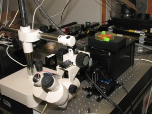
A new high-tech microscope developed by Ernst Stelzer’s group at EMBL Heidelberg will be made available to labs everywhere in the next few years. EMBL and technology giant Carl Zeiss have signed a licensing deal to commercialise the new technology, called SPIM (for Selective Plane Illumination Microscopy).
Ernst and his group developed the SPIM technology to allow scientists at EMBL to observe complex, three-dimensional processes in whole, living organisms.
We were extremely pleased to have found Carl Zeiss as an excellent partner to translate this technology into a product.

“Especially today, it’s important to study biological systems in their real context and to move away from ‘flat’ cell biology,” Ernst says. “That’s now possible thanks to the many methods available for fluorescent microscopy. The technical innovations we have introduced, including software developed by Jim Swoger to combine multiple images, will certainly be of great help.”

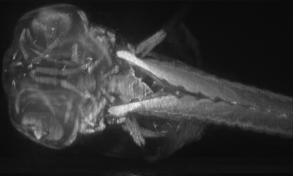
With SPIM, a sample is passed through an extremely thin sheet of light, capturing high-quality images layer by layer.
“The sample is kept alive in a liquid-filled chamber and can be rotated and viewed along different directions,” says PhD student Jan Huisken. “This eliminates blurry and unwanted light, which prevented scientists from looking deep into tissues in the past.”
Image slices are captured and quickly assembled into a high-resolution, three-dimensional film. Because light enters the sample from the side (rather than the line of sight), it is possible to achieve high resolution in all three dimensions. Illuminating the sample from the same direction as the eye or camera, as was done in the past, meant that the image was blurry along that axis.
Ernst and his colleagues are developing fascinating applications of the instrument with Jochen Wittbrodt and other EMBL scientists. The presentation of SPIM at scientific conferences has generated a flood of requests for the instrument. Last month, EMBL Director General Fotis C. Kafatos, EMBLEM Deputy Managing Director Martin Raditsch, and Ernst met with Carl Zeiss’ Executive Board Member Norbert Gorny and General Manager for Microscopy Ulrich Simon to finalise the details of the agreement.
“We were extremely pleased to have found Carl Zeiss as an excellent partner to translate this technology into a product,” says Martin. Carl Zeiss is equally happy with the agreement.
According to Ulrich Simon, “The products based on this technology will form a perfect match with our lines of confocal and multi-photo 3D-imaging systems.”

This article was originally published in EMBLetc., issue 26, April 2005. Find more stories about EMBL and SPIM here.
Related links
EMBL and ZEISS enter long-term strategic partnership