Meet the poster prize winners of ‘Cellular mechanisms driven by phase separation’
For many attendees of the EMBO | EMBL Symposium ‘Cellular mechanisms driven by phase separation’, it was the first in-person event after a long while. The excitement was tangible all around the Advanced Training Centre, but especially along the helices where the posters were being displayed. With 108 posters to choose from, the participants had lots of fascinating science to discover and could vote for their favorite until the third day of the event. The fourteen poster presenters who received the most votes had the chance to talk to the scientific organisers who then decided on the final poster prize winners.
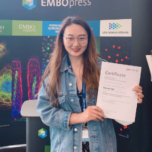
Congratulations to the winners: Gea, Alberto, Tom, and Daxiao!
A conserved mechanism regulates reversible amyloids of pyruvate kinase in yeast and human cells
Presenter: Gea Cereghetti, ETH Zürich, Switzerland

Abstract
Amyloids were long viewed as irreversible, pathological aggregates, often associated with neurodegenerative diseases. However, recent insights challenge this view, providing evidence that reversible amyloids can form upon stress conditions and fulfil crucial physiological functions. Yet, the molecular mechanisms regulating functional amyloids and the differences to their pathological counterparts remain poorly understood.
Here, we investigate the conserved principles underlying amyloid reversibility by studying the essential ATP‑producing enzyme pyruvate kinase (PK) in vitro, in yeast, and in human cells. We demonstrate that PK forms stress‑dependent reversible amyloids through a pH‑sensitive amyloid core. Stress‑induced cytosolic acidification promotes PK amyloid formation via protonation of specific glutamate (in yeast) or histidine (in human) residues within the amyloid core. Upon aggregation, PK becomes inactive and is protected from stress‑induced degradation. After stress release, re‑solubilization of yeast PK is essential to restore ATP production, disassemble stress granules (SGs), and restart cell growth. Mechanistically, we demonstrate that yeats PK re‑solubilization is initiated by the glycolytic metabolite fructose‑1,6‑bisphosphate, which directly binds PK amyloids, allowing Hsp104 and Ssa2 chaperone recruitment and aggregate re‑solubilization.
In summary, our work unravels a conserved and potentially widespread molecular mechanism underlying amyloid functionality and reversibility, and highlights the important physiological implications of regulated protein aggregation.
Fantastic discussions at the #EESPhaseSeparation poster session! It’s so stimulating to meet and discuss in person again!😀 https://t.co/yogC6iSkIo
— Gea Cereghetti (@GeaCereghetti) May 11, 2022
From Surfactant to glue – how Ki-67 regulates chromosome surface properties
Presenter: Alberto Hernández Armendáriz, EMBL Heidelberg, Germany

Abstract
Compartmentalization into functional units is a key principle of cellular life. In addition to membrane‑bound organelles, eukaryotic cells utilize membrane‑less biomolecular condensates to locally concentrate proteins and nucleic acids. While we are beginning to understand how membrane‑less condensates assemble and disassemble, we know very little about the biological processes that take place at the surface of such condensates. The surface of the largest membrane‑less cellular assembly, the mitotic chromosome, is covered by the intrinsically disordered protein Ki‑67. Our previous studies have revealed that Ki‑67 has dual functionality. In early mitosis, Ki‑67 functions as a surfactant to prevent chromosomes from collapsing into a single chromatin mass, whereas it actively promotes chromosome clustering during exit from mitosis. How Ki‑67 switches between these two opposing processes – chromosome dispersal and chromosome clustering – has remained unknown. Here, we demonstrate that Ki‑67’s biophysical properties radically change during anaphase onset, when all chromosomes merge into a single cluster. Ki‑67’s amphiphilic character is lost as its molecular brush structure collapses and the soluble pool of the protein forms condensates. Our study uncovers a cell‑cycle‑regulated mechanism that controls individualization and coalescence of chromosomes during mitosis.
Due to data protection regulations, we cannot publish the poster.
Resolving molecular ageing processes of nuclear pore proteins using a microfluidic droplet assay
Presenter: Tom Scheidt, University of Mainz, Germany

Abstract
The physiological permeability barrier for molecular traffic between the nucleus and the cytosol is filled with intrinsically disorder proteins (IDPs) and assembled by the nuclear pore complex (NPC). These highly enriched disordered nuclear proteins contain domains with high amounts of phenylalanine and glycine (FG‑Nups). In order to understand the physico‑chemical properties of such molecular “gatekeeper”, we made use of a microfluidic device capable of controlled protein condensation, combined with fast and parallelised data acquisition. Our microfluidic device permits studying phase separation on the seconds time scale (due to diffusive mixing and laminar flow), coupled with rapid optical inspection of permeability barrier properties. This time resolution is challenging to achieve by conventional benchtop experiments such as coverslip assays. Our experiments show a rapid aging of FG‑Nups into different material states (liquid, gel, solid etc.) within minutes under physiologically relevant concentrations. Already early droplets show typical properties of a liquid state and resemble a barrier and cargo delivery properties found for physiological nuclear transport. This includes formation of a natural barrier for cargoes larger than ~4 nm, unless accompanied by nuclear transport receptors (NTRs). For a better understanding of the evolution of supramolecular structures as well as the mechanical properties of FG‑rich droplets, we combine our microfluidic system together with coherent anti‑Stokes Raman spectroscopy (CARS) and particle tracking microrheology (PTM). This interdisciplinary approach provides a coherent picture to explain how the balance between homo‑ and heterotypic interactions in FG/NTR mixtures modulates the phase behavior and how this relates to the permeability barrier function of the condensed liquid state. The microfluidic platform described above can work as a general tool to study LLPS of phase separating proteins, particularly those that undergo rapid maturation to gel or amyloid like states, such as e.g. FUS, Tau and alpha‑synuclein or other proteins associated to neurodegeneration.
Reconstitution of tight junction like networks via ZO1 surface condensation and local actin polymerization
Presenter: Daxiao Sun, Max Planck Institute, Germany

Abstract
Tight junctions are adhesion structures of cell-cell contact in epithelial and endothelial. Biochemical and structural analysis revealed that tight junctions are composed of densely packed proteins forming a continuous membrane associated network at sub-apical, and are involved in adhesion, barrier, polarity, development and mechanotransduction functions, although the molecular basis underlying the assembly and positioning of tight junction network is not clear. Here we combined a bottom-up reconstitution approach on SLBs and computational simulation to investigate the mechanism behind tight junction network formation. We found that tight junction scaffold protein zona occludens1 (ZO1) undergoes surface phase separation with receptor proteins on SLBs and forms membrane condensates under a physiological concentration which is far below its 3D saturation concentration. We also showed that this process depends on receptor valency, receptor density, and ZO1 concentration. With AFM, we found that ZO1 membrane condensates are 2D structures with a height of one-layer molecules. Moreover, these 2D membrane condensates are sufficient to recruit other tight junction related components, like ZO2, ZO3, afadin, cingulin and especially actin. The enrichment of actin to ZO1 membrane condensates promotes actin polymerization and actin bundle formation. ZO1 membrane condensates deform on actin bundles simultaneously, and eventually form a continuous receptor-ZO1-actin network on SLBs together. Applying computational simulation, we showed surface phase separation with the presence of specific surface binding under saturation concentration, and the dependence on receptor valency, receptor density and ZO1 bulk concentration. Thus, combining in vitro reconstitution and computational simulation, our results suggest that surface phase separation and local actin polymerization underlies tight junction network formation. And this approach and mechanism could be applied to investigate and explain other membrane associated compartments formation.
Due to data protection regulations, we cannot publish the poster.
The EMBO | EMBL Symposium ‘Cellular mechanisms driven by phase separation’ took place from 9 – 12 May 2022.