Best poster prizes at ‘Transcription and chromatin’
The EMBL Conference ‘Transcription and chromatin‘ took place last month at EMBL Heidelberg. This popular meeting has a long-standing tradition in shaping the field of transcriptional regulation. The conference brings together leading experts covering all aspects of transcription including cis-regulatory function, long range regulation, 3-dimensional looping, the basal transcriptional machinery, RNA polymerase regulation and function, nucleosome positioning, chromatin modifications, chromatin remodelling, and epigenetic inheritance of transcriptional silencing.
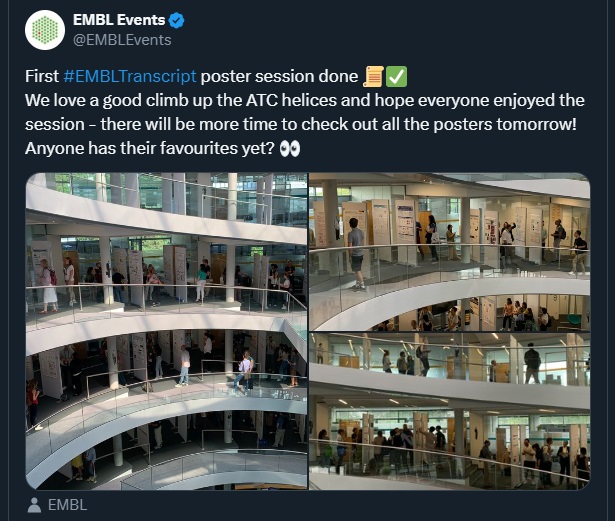
For the 16th edition of this meeting, we had 347 people attending on-site and 100 virtual participants. There were nineteen fellowships provided by the EMBL Corporate Partnership Programme and EMBO. With the total of 262 posters to view, we held three poster sessions during which the presenters could discuss their research — their work was then voted for by the poster committees formed by selected participants with the support of our scientific organisers. There were five poster prizes awarded during the meeting – two of them kindly sponsored by FEBS Letters. We are pleased to share with you all of the awarded abstracts!
Irregular nucleosome spacing drives chromatin into a more open and dynamic state
Presenter: Waad AlBawardi
Authors: Waad AlBawardi, Matthew Thomas, Gert-Jan Kuijntjes, Jim Allan, Willem Vanderlinden, Chris A Brackley, John van Noort, Nick Gilbert, Hannah Wheeldon

Abstract:
Chromatin is the substrate for all DNA associated processes; and revealing its structure is key to understanding the regulation of nuclear functions. Accumulating evidence suggests that chromatin adopts variable structures inside cells, yet the molecular mechanisms for regulating chromatin structure remain elusive. Nucleosome positioning patterns vary by genomic regions, with constitutive heterochromatin domains having regularly positioned nucleosome arrays, unlike the variable positioning in euchromatin. How nucleosome positioning irregularity affects chromatin structure and mechanical properties is poorly understood. To address this question, we synthesized novel physiologically relevant templates that contain nucleosome positioning sequences derived from the ovine β lactoglobulin (BLG) gene. The templates have variable nucleosome positioning analogous to positioning patterns in active euchromatin. The Widom 601 template, which is composed of tandem repeats of the 601 motif that forms strongly positioned nucleosomes, is used to model heterochromatic regions.
We show that irregular nucleosome arrays form heterogeneous and disrupted structures when folded in the presence of the linker histone H5. Analysis of the dynamic properties of fibres using polymer modelling and magnetic tweezers shows that irregular fibres are more dynamic and fragile under tension compared to uniform fibres. Together, these findings indicate that intrinsic nucleosome regularity may enable heterochromatin to maintain a stable compact state, facilitating gene silencing. In contrast, variable positioning provides dynamic plasticity to open and remodel euchromatic regions. This data also reconciles major discrepancies between classical ordered models derived from in vitro studies with repetitive sequences and recent in situ evidence for heterogeneous chromatin fibres in living cells.
Due to the confidentiality of the unpublished data, we cannot share the poster.
Escape from X inactivation is directly modulated by Xist RNA levels
Presenter: Agnese Loda
Authors: Antonia Hauth, Jasper Panten, Emma Kneuss, Christel Picard, Nicolas Servant, Isabell Rall, Yuvia A. Pérez Rico, Lena Clerquin, Nila Servaas, Laura Villacorta, Ferris Jung, Christy Luong, Howard Chang, Oliver Stegle, Duncan Odom, Agnese Loda, Edith Heard

Abstract:
In placental females, one copy of the two X chromosomes is largely silenced during a narrow developmental time window, in a process mediated by the non coding RNA Xist. Here, we demonstrate that Xist can initiate X chromosome inactivation (XCI) well beyond early embryogenesis. By modifying its endogenous level, we show that Xist has the capacity to actively silence genes that escape XCI both in neuronal progenitor cells (NPCs) and in vivo, in mouse embryos. We also show that Xist plays a direct role in eliminating TAD like structures associated with clusters of escapee genes on the inactive X chromosome, and that this is dependent on Xist’s XCI initiation partner, SPEN. We further demonstrate that Xist’s function in suppressing gene expression of escapees and topological domain formation is reversible for up to seven days post induction, but that sustained Xist up regulation leads to progressively irreversible silencing and CpG island DNA methylation of facultative escapees. Thus, the distinctive transcriptional and regulatory topologies of the silenced X chromosome is actively, directly and reversibly controlled by Xist RNA throughout life.
Structural insights into transcription elongation on chromatin in cells
Presenter: Tomoya Kujirai
Authors: Tomoya Kujirai, Haruhiko Ehara, Junko Kato, Kyoka Yamamoto, Seiya Hirai, Takeru Fujii, Kazumitsu Maehara, Akihito Harada, Lumi Negishi, Mitsuo Ogasawara, Mikako Shirouzu, Yuki Yamaguchi, Yasuyuki Ohkawa, Yoshimasa Takizawa, Shun ichi Sekine, Hitoshi Kurumizaka

Abstract:
In eukaryotic cells, the genomic DNA forms the chromatin structure, in which the nucleosome is the basic unit. RNA polymerase II (RNAPII) is a central enzyme to produce messenger and non coding RNAs. RNAPII must transcribe the DNA tightly wrapped around the histone proteins. Transcription elongation factors and histone chaperones are involved in chromatin transcription. Using an in vitro nucleosome transcription system, we have analyzed the effects of the transcription elongation factors and the histone chaperone FACT and determined the structures of the transcribing RNAPII elongation complex nucleosome FACT by cryo electron microscopy (cryoEM). These structures reveal nucleosome disassembly/reassembly with the aid of the transcription elongation factors and the histone chaperone.
To gain deeper insight into chromatin transcription in vivo, we have conducted an ex vivo approach, ChIP cryoEM method. In this method, chromatin of the HeLa cells expressing FLAG tagged RPB3, an RNAPII subunit, was digested with micrococcal nuclease. The RNAPII complexes are purified by chromatin immunoprecipitation (ChIP), followed by sucrose gradient fixation. We then determined the structures of RNAPII molecules transcribing the genomic DNA with/without endogenous transcription elongation factors, SPT5, SPT6, and ELOF1. In addition, the structures of transcribing RNAPII with an endogenous nucleosome were determined. In the RNAPII downstream nucleosome, RNAPII paused at the SHL( 5) position on the nucleosome, consistent with previous genome wide analysis (Weber et al., Mol Cell, 2014). We found an RNAPII upstream nucleosome complex, in which the histone H2A H2B in the nucleosome directly contacts the RNAPII surface, suggesting that RNAPII pauses after nucleosome reassembly. These structures may represent the nucleosome disassembly/reassembly process in cells.
In this meeting, we will discuss the mechanisms of chromatin transcription from the structural perspective.
Due to the confidentiality of the unpublished data, we cannot share the poster.
Catalytic and non-catalytic functions of p300/CBP in zygotic genome activation
Presenter: Sergei Pirogov
Authors: Sergei Pirogov, Mattias Mannervik, Melissa Harrison, Audrey Marsh, Suresh Sajwan

Abstract:
The p300/CBP acetyltransferase is a co activator of gene expression that establishes the H3K27ac histone mark on active enhancers and promoters. The exact mechanism by which CBP mediated acetylation promotes transcription remains elusive. Previous studies have shown that while substitution of H3K27 using histone replacement does not affect gene activation, chemical inhibition of CBP in cell culture affects transcription initiation and pause release of many genes. We sought to reveal the mechanisms by which CBP functions in vivo using genetic approaches during zygotic genome activation (ZGA) in Drosophila melanogaster. To inactivate endogenous CBP in the zygote, we used two orthogonal approaches: loading of embryos with a catalytically dead CBP, or optogenetic inactivation of CRY2 tagged CBP. In both of these cases, CBP catalytic activity was disabled while CBP protein levels remained intact. We compared these embryos to those where CBP was degraded with the deGradFP system.
We found that inactivation of CBP halts gastrulation due to downregulation of many zygotic genes. To explore the mechanism of downregulation, we performed CUT&Tag and qPRO seq to measure the distribution of CBP and RNA polymerase II (Pol II). After rapid CBP inactivation with the optogenetic approach, Pol II accumulated on zygotic gene promoters but was reduced in gene bodies, suggesting a pause release defect. When CBP was degraded, we observed a decrease of Pol II on both promoters and gene bodies, suggesting a non catalytic CBP function in Pol II recruitment. In embryos with catalytic dead CBP, the effect was blended since nearly half of the CBP peaks had reduced CBP occupancy. The Zelda (Zld) motif was enriched at these peaks, while regions with GAGA factor (GAF) enriched motifs did not lose CBP. Based on these results, we propose a model where GAF recruits inactive CBP to promoters of highly paused genes to promote initiation and pausing of Pol II in a non catalytic manner. By contrast, CBP activity is needed for interaction with Zld bound enhancers, which stimulates pause release in a catalytic dependent manner.
Poster prize kindly sponsored by FEBS Letters.
Distinct mechanisms of chromatin engagement by transcriptional activators and repressors: insights from bHLH transcription factors
Presenter: Lisa Stoos
Authors: Lukas Kater, Lisa Stoos, Georg Kempf, Simone Cavadini, Fumiya Moribe, Nikolas Eggers, Dirk Schübeler, Nicolas Thomä

Abstract:
Eukaryotic DNA is tightly organized into nucleosomes, rendering it difficult for chromatin binding factors, including transcription factors (TFs), to engage specific DNA sites on nucleosomes. The basic helix loop helix (bHLH) family, the third largest family of TFs, recognizes E box motifs (CANNTG). Only a fraction of E box motifs in the genome are occupied at any time. Our previous study explored how bHLH members c MYC MAX (bHLH LZ) and CLOCK BMAL1 (bHLH PAS) bind to their target sites within nucleosomes, revealing general principles of chromatin engagement.1 Both preferentially bind E boxes near the nucleosomal entry exit sites. Structural studies show that MYC MAX and CLOCK BMAL1 trigger DNA release from histones to gain access. The TFs stabilize this release through histone contacts via auxiliary domains, such as dimerization domains. CLOCK BMAL1 utilizes flexible linkers to optimize histone contacts, while we find MYC MAX, with a rigid leucine zipper, engaging histones predominantly in histone facing motif positions. Recent unpublished structural and functional investigations into the repressor MAD1 MAX revealed a unique nucleosome binding mechanism distinct from MYC MAX. MAD1 MAX targets E boxes at the nucleosomal entry exit site and linker, and when doing so, dimerizes adjacent nucleosomes through its leucine zipper domain. Detailed examination of the structures identified three arginine anchors crucial for nucleosome dimerization. Similar interacting residues are evident in other MAX binding partners that also serve as repressors. Functional implications of the different nucleosome binding modes for repressive vs. activating TFs of the same fold family will be discussed.
Due to the confidentiality of the unpublished data, we cannot share the poster.
Poster prize kindly sponsored by FEBS Letters.

The EMBL Conference ‘Transcription and chromatin’ took place from 24 – 27 August 2024 at EMBL Heidelberg and virtually.