Best poster prizes at ‘Defining and defeating metastasis’
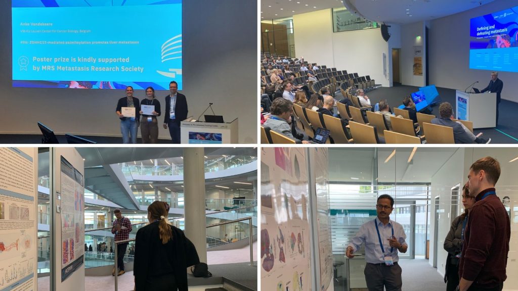
The EMBO | EMBL Symposium ‘Defining and defeating metastasis‘ took place last month at EMBL Heidelberg. In its second edition, this symposium covered emerging key concepts of latent metastasis and dissemination of tumour cells including evolution and genetic diversity of metastasis. With 150 onsite participants, 70 virtual participants, and 14 fellowships provided by the ‘EMBL Corporate Partnership Programme‘ and ‘EMBO‘, we had the opportunity to gather metastasis researchers, PhD students, and experts in the field form different parts of the world. The event provided a unique interdisciplinary exchange on current approaches and future collaborations on metastasis and its therapeutic challenges. With a total of 104 posters available, we held two poster sessions during which the presenters could discuss their research — their work was then voted by the participants. There were four poster prizes awarded during the meeting.
Please give a round of applause to these brilliant ladies!
Zeb1 expression in cancer-associated fibroblasts promotes distant metastasis in PDAC
Presenter: Elisabetta D’Avanzo
Authors: Elisabetta D’Avanzo, Isabell Armstark, Florian Krämer, Harald Schuhwerk, Pooja Gupta, Ruthger van Roey, Kathrin Fuchs, Dieter Saur, Thomas Brabletz, NathalieBritzen-Laurent, Marc P. Stemmler
Poster prize is kindly supported the EMBO Journal

Abstract:
Most patients with pancreatic ductal adenocarcinoma (PDAC) rapidly succumb the disease due to late prognosis and early metastasis. PDAC is very dysplastic, containing up to 80% stroma, which is mainly consisting of cancer-associated fibroblasts (CAFs). Besides its well-studied function in tumor cell invasion, metastasis and stemness, the transcription factor Zeb1 is also frequently upregulated in the stroma and increased stroma Zeb1 expression has been correlated with poor patient’s survival. We here addressed the role of Zeb1 in CAFs for tumor progression. Our previous analyses have demonstrated that depletion of Zeb1 in CAFs (FibΔZeb1) of colorectal cancer (CRC) affects CAF diversification with impaired myCAF and altered iCAF function that results in increased immune cell infiltration, beneficially increasing sensitivity to immunotherapy. Here we show that the inactivation of Zeb1 in Col6+ fibroblasts in an autochthonous PDAC mouse model does not affect primary tumor growth and differentiation but strongly reduces metastasis. Since experimental colonization to lungs and liver did not change upon i.v. injection of tumor cells, we are focusing on events, which depend on the primary tumor to alter metastasis in FibΔZeb1 mice, including systemic premetastatic niche priming events. scRNAseq and functional analyses of CAFs from PDAC shows that Zeb1-depleted CAFs are less diverse and exhibit a loss of antigen-presenting CAFs (apCAFs), reduced myCAFs and modified iCAFs features, including an increase in cytokine secretion. In contrast to the CRC model, no general increase in immune cell infiltration in PDAC FibΔZeb1 tumors was observed, but a detailed characterization that focuses on the immune suppressing function of apCAFs is ongoing to dissect how CAFs in the primary tumor regulate metastasis. Our data show that CAF specification and abundance of subpopulations play an important role for tumor progression and metastasis and interference with CAF specification could be of therapeutic use in PDAC.
Due to the confidentiality of the unpublished data, we cannot share the poster.
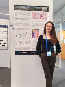
Transcriptional patterns of primary and metastatic NSCLC
Presenter: Alexandra Beno
Authors: Alexandra Beno, Kolos Nemes, Eva Mago, Petronella Topolcsányi, Gabriella Mihalekné Fur, Lorinc S. Pongor
Poster prize kindly supported by EMBO Molecular Medicine

Abstract:
Small cell lung cancer (SCLC) is a highly aggressive malignancy characterized by rapid growth, early metastasis, and a poor prognosis. Understanding the mechanisms driving metastasis is crucial for developing targeted therapies to improve patient outcomes. Recent advancements in single-cell RNA sequencing (scRNA-seq) provide an unprecedented opportunity to dissect the transcriptional landscape of SCLC at a single-cell resolution. This study focuses on elucidating the transcriptional differences between primary and metastatic SCLC tumors, leveraging scRNA-seq to identify key markers and regulatory pathways involved in metastasis. We investigate the transcriptional control of metastasis by analyzing the expression of critical transcription factors, signaling pathways, and metastasis-associated genes. Heterogeneity within metastatic tumors is explored through clustering, revealing distinct subpopulations. The role of epithelial-mesenchymal transition (EMT) in SCLC metastasis is assessed by evaluating the expression of various epithelial and mesenchymal markers. We characterize tumor-immune interactions by identifying and annotating various immune cell populations, analyzing the expression of immune checkpoint molecules, and inferring ligand-receptor interactions. By integrating comprehensive analyses, we aim to uncover the transcriptional drivers of SCLC metastasis, the heterogeneity within metastatic tumors, the EMT process, and mechanisms of immune evasion. Our findings will contribute to a deeper understanding of SCLC biology and may inform the development of novel therapeutic strategies targeting metastatic disease.
Leveraging 3D biomimetic in vitro models to dissect the bone metastatic cascade from breast cancer
Presenter: Chiara Spadazzi
Authors: Chiara Spadazzi, Claudia Cocchi, Sofia Gabellone, Silvia Vanni, Alessandro De Vita, Giacomo Miserocchi, Chiara Calabrese, Sofia Dellavalle, Oneda Cani, Lucas Moron, Dalla Tor, Michela Tebaldi , Chiara Liverani
Poster prize is kindly supported by MRS Metastasis Research Society

Abstract:
Background Bone metastasis is a debilitating and incurable disease occurring in
approximately 70% of metastatic breast cancer cases. The intricate interactions between
tumor cells and the bone microenvironment (BME) govern each step of the metastatic
journey, providing windows of opportunities for therapeutic intervention. Here, we aimed to
leverage 3D biomimetic models to mimic the bone metastatic process in vitro.
Methods Two breast cancer (BC) cell lines, MDA-MB-231 (triple-negative BC) and MCF7 (luminal A BC), were used. Breast tissue and bone-specific ECM were mimicked with a type I collagen-based scaffold and a collagen hydroxyapatite (HA) scaffold. RNA Seq analysis was employed to evaluate the ECM-modulation in the BC tumor-subtypes. The bonemimetic scaffold was cellularized with osteoclast progenitor cells, optimized for differentiation. A bone metastasis (BM) model was settled up combining scaffolds together to mimic the distant metastatic process in vitro for 14 days. Then, cells were characterized
by confocal imaging and qRT-PCR.
Results RNA Seq showed BC cells modulated different pathways: hypoxia and JAK-STAT for MDA-MB-231; MAPK signaling and lipid metabolism for MCF7. In our BM model, MCF7 co-cultured with osteoclasts upregulated stemness and mesenchymal related-markers (CD44, TGF-beta, VIM, ZEB1, ZEB2). MDA-MB-231 downregulated mesenchymal markers, suggesting a mesenchymal to epithelial transition induced by bone-ECM. Moreover, these adaptations were morphologically confirmed by confocal imaging. Besides different mechanisms, both cell lines induced osteoclast differentiation, demonstrated by OC markers upregulation (CA2, OSCAR, RANK).
Conclusions The model enables us to parse apart the metastatic process from BC,
highlighting subtype-specific mechanisms governing establishment of metastasis. This
model could aid the identification of potential therapeutic strategies that specifically targetdefined steps to prevent or treat skeletal disease.
Due to the confidentiality of the unpublished data, we cannot share the poster.
ZDHHC17-mediated palmitoylation promotes liver metastases
Presenter: Anke Vandekeere
Autors: Anke Vandekeere , Sarah-Maria Fendt
Poster prize is kindly supported by MRS Metastasis Research Society

Abstract:
Metastasis formation is the leading cause of mortality in breast cancer patients. The liver, one of the most common sites of metastases, has recently been identified as an environment rich in lipids such as palmitate and oleate. Also, further increasing palmitate abundance through high fat diet was shown to induce more liver metastases. However, it is still not completely understood how cancer cells use this surrounding palmitate to boost their metastatic growth in the liver. Interestingly, we found that breast cancer cells that metastasize in the liver use the high levels of environmental palmitate to post-translationally modify proteins through palmitoyl-transferase ZDHHC17 and hereby promote a pro-tumoral microenvironment. We found that reducing ZDHHC17 expression in the cancer cells, and subsequently removing the lipid-derived protein modification on specific proteins, can decrease liver metastases by redirecting the surrounding stromal cells towards an anti-tumoral response. Indeed, the loss of ZDHHC17 in cancer cells creates a tumor microenvironment in which the cancer cells are killed by surrounding cells – a phenotype that can be reversed by depleting or inhibiting specific cell functions. In conclusion, our data shows that the high palmitate abundance in the liver fosters metastatic growth by modifying the communication in the tumor microenvironment. Targeting the lipid-derived modification of these signals could allow us to rewire the surrounding cells towards an anti-tumoral response and opens new therapeutic strategies for liver metastases.
Due to the confidentiality of the unpublished data, we cannot share the poster

The EMBO | EMBL Symposium ‘Defining and defeating metastasis’ took place from 30 September – 3 October 2024 at EMBL Heidelberg and virtually.