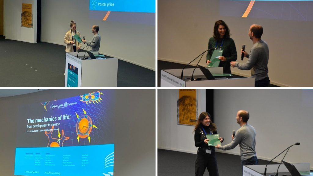
Best poster prize winners of the EMBO | EMBL Symposium ‘The mechanics of life: from development to disease’
Last April, our EMBO | EMBL Symposium ‘The mechanics of life: from development to disease’ took place in Heidelberg, Germany and virtually. We were happy to welcome 173 attendees to the Advance Training Centre in Heidelberg, Germany, and 57 virtual participants. This event gathered top experts to explore the interplay of the physical forces between the fundamental building blocks of life, from molecules to organs.
In addition to the talks, 98 researchers presented their work at the poster sessions. Participants voted for the best posters and today we present you the winners. Let’s give a round of applause to celebrate the extraordinary contributions of our poster prize winners.
Congratulations to all the winners!
Mechanics of cardiomyocyte nuclei in a beating heart
Presenter: Laura Bader
Collaborators: Marie-Christine Ramel and Rashmi Priya

Abstract:
During development, cells experience diverse mechanical forces and adapt by changing their shape, behaviour or eliciting a specific signalling response. How the nucleus, which is one of the largest and most rigid organelles inside a cell responds to mechanical forces in a developing tissue during embryonic development remains understudied. The vertebrate heart is a highly mechanically active organ, as it generates substantial forces during each heartbeat. How cardiomyocyte nuclei respond to these forces and sustain their homeostasis in a live beating heart remain less understood. To address this question, here we have used the developing zebrafish heart as a model system combined with multiscale imaging, morphometrics, chemical and genetic perturbations. Interestingly, cardiomyocytes exhibit a distinct nuclear morphology depending on their spatial localisation – taut, homogeneously shaped nuclei in atrium and variable nuclear morphology in ventricle; with the outer enveloping compact layer showing a variable lobular morphology while the inner contractile trabecular layer has more taut nuclei. Of note, manipulation of cardiac forces alters nuclear morphology in the ventricle in a dose dependent manner. Decreasing forces lead to rounder and taut nuclei with decreased volume while increasing forces lead to lobular nuclear morphology and increased volume. Further, initial data indicates enrichment of microtubules in a cage-like structure in proximity to cardiomyocyte nuclei, and alteration of nuclear shape following experimental microtubule disassembly. We are currently following up on these observations to understand why cardiomyocyte nuclei have different morphology in different tissue layers and how microtubules and nuclear lamins contribute to these morphological differences. We believe that these results will expand our knowledge of nuclear mechanosensing and improve our understanding of how nuclear homeostasis is sustained in a mechanically dynamic tissue during development.
Due to the confidentiality of the unpublished data, we cannot share the poster.
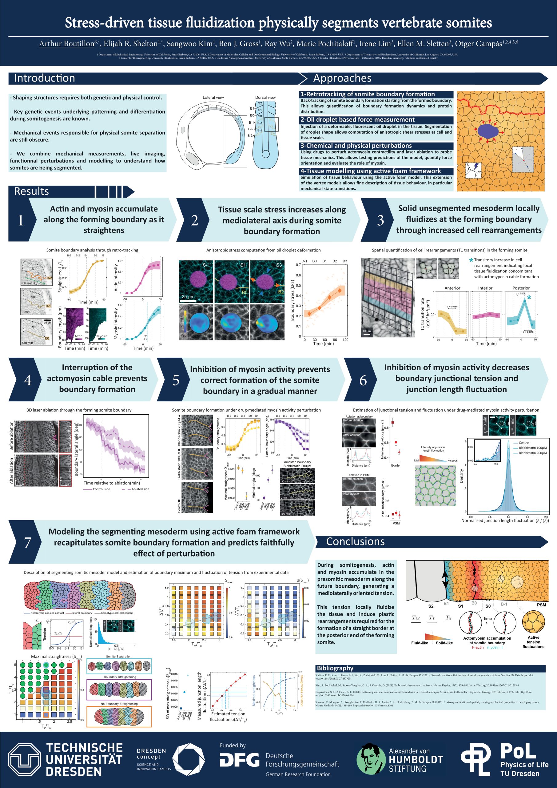
Stress-driven tissue fluidization physically segments vertebrate somites
Presenter: Arthur Boutillon
Collaborators: Elijah Shelton, Sangwoo Kim, and Otger Campàs

Abstract:
Shaping functional structures during embryonic development requires both genetic and physical control. During somitogenesis, cell-cell coordination sets up genetic traveling waves in the presomitic mesoderm that orchestrate somite formation. While key molecular and genetic aspects of this process are known, the mechanical events required to physically segment somites from the presomitic mesoderm remain unclear. Combining direct mechanical measurements during somite formation, live imaging of cell and tissue structure, pharmaceutical and mechanical perturbations and computer simulations, we show that somites are mechanically sectioned off from the presomitic mesoderm by a large, actomyosin-driven increase in anisotropic stress at the nascent somite-somite boundary. Our results show that this localized increase in tensile stress drives the regional fluidization of the tissue adjacent to the forming somite border, enabling local tissue remodeling and the shaping of the somite. We observed that myosin inhibition decreases tension in the forming boundary, resulting in failure to completely separate the somites. Moreover, we find that active tension fluctuations in the tissue are optimized to mechanically define sharp somite boundaries while minimizing somite morphological defects. Altogether, these results indicate that mechanical changes at the somite-somite border and optimal tension fluctuations in the tissue are essential physical aspects of somite formation.
ECM remodelling and tension development in tendon wound healing is mechanically regulated
Presenter: Alina Dintheer
Collaborators: Amro Hussien, Patrick Jaeger, and Jess Snedeker

Abstract:
Background: ECM mechanical cues guide healing in tendons. Yet, the molecular mechanisms orchestrating healing processes remain elusive. Appropriate tissue tension is essential for tendon homeostasis and tissue health. By mapping the attainment of tissue-level tensional forces, we aim to understand how ECM tension regulates tendon healing. We hypothesize that diseased tendon returns to homeostasis only after the cells have reached a mechanically gated exit from wound healing. Methods: We engineered a 3D mechano-culture system to create tensioned tendon-like constructs by embedding patient-derived tendon cells into a collagen I hydrogel. Casting the hydrogel between posts anchored in silicone allowed adjusting the post stiffness. Under this static mechanical stimulation, cells remodel the (unorganized) collagen representing wound healing mechanisms. We quantified the tissue-level forces using post deflection measurements. Newly deposited ECM was visualized by metabolic labelling with non-canonical amino acids, click chemistry and confocal microscopy. Relative mRNA expression of ECM genes was assessed by RT-qPCR. We inhibited the tension development by blocking actin-myosin contractility using a ROCK inhibitor (Y27632). Results: We found that cell-mediated tensional forces of microtissues cultured on stiffer posts (k = 5N/m) reached higher levels compared to compliant controls (k = 1N/m, p < 0.0001). While minimal matrix was synthesized in early phases of tissue formation (d3-d5), cell-deposited ECM was present in later stages (d7-d9). More ECM was deposited by tendon constructs cultured on compliant compared to rigid posts (p = 0.0017), along with significantly downregulated ECM genes Col1a1, Col3a1, FN, LOX. Matrix synthesized by low-tensioned constructs was less aligned indicated by greater dispersion of fibers (p = 0.0021). Inhibiting ROCK decreased tissue-level tensional forces (p < 0.0001). Conclusions/Outlook: Our results indicate that tendon cells balance matrix remodelling and synthesis during the different phases of tissue repair reaching tension levels and matrix status as a function of the mechanical microenvironment. We are identifying molecular mechanosensors and regulatory pathways guiding tension-mediated exit from wound healing towards homeostasis.
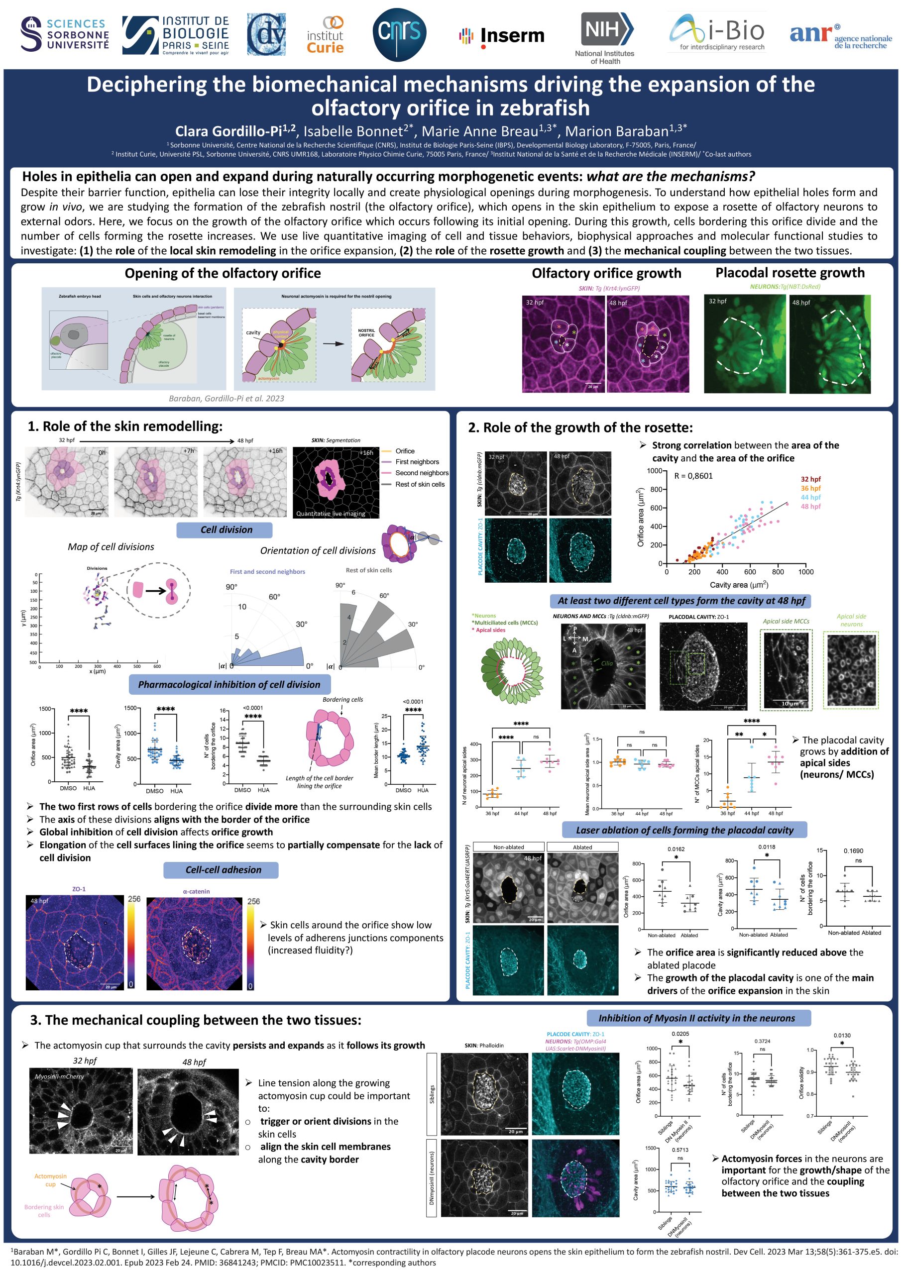
Deciphering the biomechanical mechanisms driving the expansion of the olfactory orifice in zebrafish
Presenter: Clara Gordillo-Pi
Collaborators: Isabelle Bonnet, Marie Breau, and Marion Baraban

Abstract:
Despite their barrier function, epithelia can lose their integrity locally and create physiological openings during morphogenesis. To understand how epithelial holes grow in vivo, we are studying the formation of the zebrafish nostril (the olfactory orifice), which opens in the skin epithelium to expose olfactory neurons to external odors. We have previously shown that a subset of olfactory neurons assemble into a rosette that opens the olfactory orifice by pulling on the overlying skin cells. By analyzing fixed samples, we noticed that after opening, the olfactory orifice continues to grow in the skin, a process that is concomitant with an increase in both the number of skin cells bordering the orifice and the number of cells forming the rosette. Using live quantitative imaging, we showed that the skin cells lining the growing orifice divide more than the surrounding skin cells and do so tangentially to the edges of the growing orifice, and that the underlying rosette develops through the intercalation of new cells, coming from the basal region of the olfactory placode. Pharmacological inhibition of proliferation in the whole embryo decreased the size of the olfactory orifice, suggesting that cell divisions in the bordering cells are necessary for the orifice expansion. In addition, specific perturbations in rosette cell number led to a smaller orifice in the skin, indicating that neuronal rosette growth is a major contributor to the orifice expansion in the skin epithelium. Finally, inhibition of Myosin-II and mechanosensitive channels revealed a mechanical coupling between the two tissues, which appears to be necessary for the correct growth of the olfactory orifice in the skin. By combining live quantitative imaging of cell and tissue behaviors, biophysical approaches and molecular functional studies, we are further investigating the underlying mechanical and molecular mechanisms.
Stochastic and deterministic regulation of cell cycle (de)synchronization ensures robust initiation of embryo morphogenesis
Presenter: Magdalena Schindler
Collaborators: Daniel R. Amor, Bernat Corominas-Murtra, and Nicoletta Petridou

Abstract:
A conserved process of early embryonic development in metazoans is the reductive cell divisions following oocyte fertilization, termed cell cleavages. Cell cleavages usually desynchronize before major morphogenetic events. Although cell cycle dynamics are regulated at the single cell level, the cell-to-cell desynchronization observed in early embryos elicits collective effects, including shape and pattern formation. This raises the question of how single-cell controllers coordinate embryo-scale processes. Here, we address this question in the zebrafish embryo, where cell cleavages were shown to impact the tissue material state, and subsequently the onset of the first morphogenetic event, blastoderm spreading. By using live imaging and quantitative data analysis we detect a super-exponential increase of cell cycle duration associated with a fast increase in cell-to-cell variability in cell cycle lengths. These rapid dynamics can be modelled as a hyperbolic growth, suggesting that the system slows down reaching a “critical” point where it would presumably arrest. Intriguingly, the embryo becomes maximally variable concerning its cell cycle, when approaching this point, coinciding with the moment of tissue fluidization. However, the embryo escapes this critical point by changing its cell cycle dynamics and drastically dropping its cell cycle variance, as the zygotic-to-embryo transition is completed. Using theory and cell biology experiments we identify that the information of cell cycle length and its noise is inherited from the mother cell and is encoded in the cell size. We propose that a balance between the generation of physically isolated cell compartments via cytokinesis and their communication via cytoplasmic bridges underlie the desynchronization dynamics to a level that ensures a robust tissue fluidization and initiation of morphogenesis. Altogether, our work highlights the importance of coordinated stochastic and deterministic mechanisms of cell cycle dynamics for regulating collective tissue properties and morphogenetic robustness.
Due to the confidentiality, we cannot share the poster.
The EMBO | EMBL Symposium ‘The mechanics of life: from development to disease‘ took place from 15 – 18 April 2024 at EMBL Heidelberg and virtually.