Lorenzo Albertazzi
Eindhoven University of Technology, Netherlands
Open access to cutting-edge electron and light microscopy
We provide researchers from Europe and beyond with a synergistic portfolio of imaging services including cryo-EM, super-resolution and intravital microscopy to enable new ground-breaking research that crosses the scales of biology.
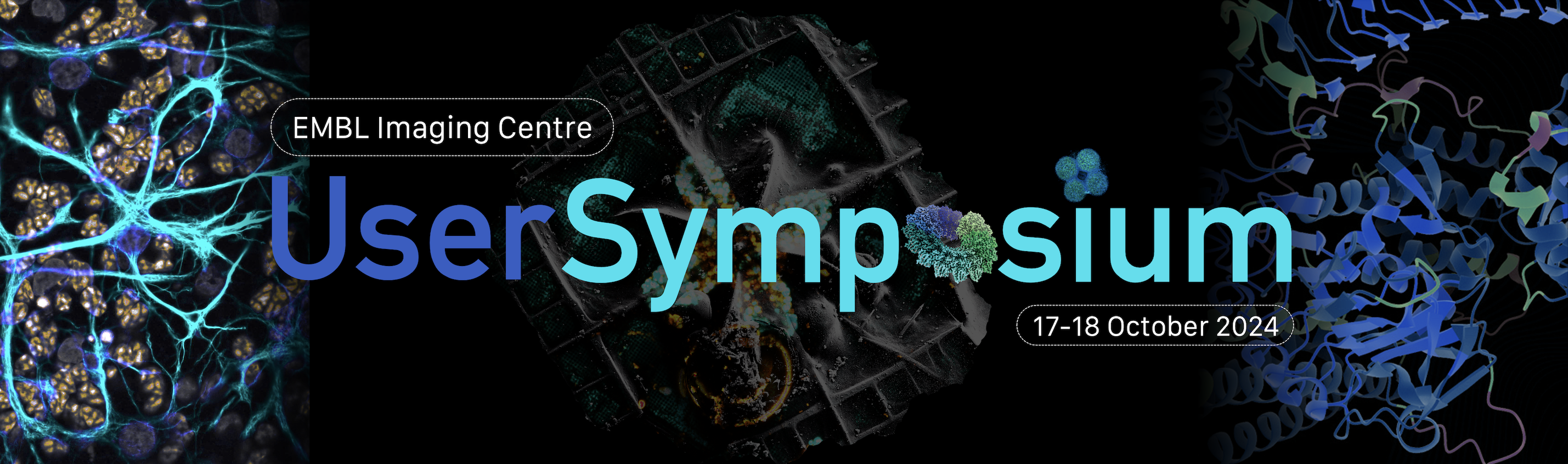
The EMBL Imaging Centre User Symposium will take place on Thursday 17th and Friday 18th October 2024, at EMBL Heidelberg, Germany, and will bring together the user community of the EMBL Imaging Centre.
The IC user symposium is covering a broad range of imaging technologies from cryo-electron and super-resolution microscopy to organismal and intravital imaging.
Invited and selected speakers have already used the facility and will present their research during the symposium. We aim to stimulate the exchange of experience among users and strengthen collaborations and interactions within the imaging community in general.
Besides the invited speakers, all participants are welcome to present their research during the poster or flash-talk session. The meeting will be rounded out with a discussion and feedback session.
Eindhoven University of Technology, Netherlands

Grenoble Alpes University, France
IGBMC, France
IGB Berlin, Germany
Delft University of Technology, Netherlands

EMBL, Germany
DKFZ, Germany

IOCB, Czech Rep.
IGBMC, France
Birkbeck University of London, UK
| Time (Europe/Berlin) | Speaker | Location |
|---|---|---|
| 12:30 – 14:00 | Registration and light refreshments | EMBL Imaging Centre registration desk |
| 13:00 – 13:45 | Industry workshops (pre-registration required) Leica: With the user, for the user – Leica Labs at the EMBL Imaging Centre supporting IC users and driving innovation in visualization and analysis (room 15-041) Zeiss: Dynamics in Life: Analyse large 4D datasets seamlessly with ZEISS arivis Cloud (room 15-104) | |
| 14:00 – 14:15 | Opening remarks Christoph Mueller – EMBL | EMBL IC Lecture Hall |
| 14:15 – 15:35 | Session 1 – Electron Microscopy Chair: Simone Mattei – EMBL | EMBL IC Lecture Hall |
| 14:15 – 14:35 | Molecular basis of mRNA delivery to the bacterial ribosome. Albert Weixlbaumer – University of Strasbourg Institute for Advanced Study, France | |
| 14:35 – 14:55 | Cryo-electron tomography reveals how COPII assembles on cargo-containing membranes. Giulia Zanetti – Birkbeck University of London, UK | |
| 14:55 – 15:15 | Decoding the cellular entry mechanism of arginine-rich peptides with a multiscale cryoEM approach Mattia Morandi – IOCB Pragu | |
| 15:15 – 15:35 | How bacteria float: Structural basis of buoyancy control with gas vesicles. Arjen Jakobi – Delft University of Technology, Netherlands | |
| 15:35 – 15:45 | Group Photo | |
| 15:45 – 16:45 | Coffee Break (sponsored by Zeiss) | EMBL IC kitchenette |
| 16:45 – 17:45 | Session 2 – CLEM Chair: Simone Mattei – EMBL | EMBL IC Lecture Hall |
| 16:45 – 17:05 | Exploring the ultrastructural organisation of a marine microalga by volume electron microscopy Johan Decelle – Grenoble Alpes University, France | |
| 17:05 – 17:25 | Linking cell architecture to genomics in the giant bacterium Achromatium. Danny Ionescu – Leibniz Institute for Freshwater Ecology and Inland Fisheries, Germany | |
| 17:25 – 17:45 | Organization of condensin proteins in the chromosomal environment in situ. Mikhail Eltsov – IGBMC, France | |
| 17:45 – 18:45 | Conference dinner | EMBL Canteen |
| 18:45 – 20:30 | Poster session / Sponsor booths Wine, dessert and cheese | EMBL IC Atrium |
| Time (Europe/Berlin) | Speaker | Location |
|---|---|---|
| 09:00 – 10:00 | Session 3 – Light Microscopy Chair: Timo Zimmermann – EMBL | EMBL IC Lecture Hall |
| 09:00 – 09:20 | Organization of crossover sites in C. elegans. Simone Köhler – EMBL | |
| 09:20 – 09:40 | Neurodevelopmental principles underlying pediatric brain cancer. Lena Kutscher – DKFZ, Germany | |
| 09:40 – 10:00 | Imaging nanoparticles, one molecule at the time. Lorenzo Albertazzi – Eindhoven University of Technology, Netherlands | |
| 10:00 – 10:45 | Coffee Break (sponsored by Zeiss) | EMBL IC kitchenette |
| 10:45 – 11:25 | Session 4 – Short Talks Chair: Timo Zimmermann – EMBL | EMBL IC Lecture Hall |
| 10:45 – 10:55 | Short Talk 1 Native molecular architectures of centrosomes in C. elegans embryos Fergus Tollervey – University of Geneva | EMBL IC Lecture Hall |
| 10:55 – 11:05 | Short Talk 2 Euro-BioImaging ERIC: Towards the democratisation of imaging technologies for scientific advancement Ayoub El Ghadraoui – Euro-BioImaging | EMBL IC Lecture Hall |
| 11:05 – 11:15 | Short Talk 3 MINFLUX tracking for monitoring stepping dynamics of kinesin-1 in live cells Takahiro Deguchi – EMBL | EMBL IC Lecture Hall |
| 11:15 – 11:25 | Short Talk 4 Structural and temporal analysis of spherical and filamentous influenza A virus cell entry and spread Sarah Peterl – University of Heidelberg | EMBL IC Lecture Hall |
| 11:30 – 12:15 | Panel Discussion Moderated by Simone Mattei and Timo Zimmermann | EMBL IC Lecture Hall |
| 12:15 – 12:45 | Closing Remarks Jan Ellenberg – EMBL | EMBL IC Lecture Hall |
Registration is for free. Places are limited.
Abstract submission deadline is 18th July 2024.
Accommodation is not included.
As a guide we have included a list of hotels you may want to consider should you require overnight accommodation:
All meals and coffee breaks are included. Our catering staff will prepare a wide variety of vegetarian meals, meat and fish dishes, soups, pasta, fresh fruit and vegetables, as well as a variety of desserts.
Please wear your badge at all times when serving yourself.
Do not smoke in any EMBL building.
Do not enter any restricted areas or the laboratories unless instructed to do so.
If first aid is required …
In case of fire …
Beyond first aid…
Please remember to bring your own medication, if needed, to the conference. Note that the next pharmacy is a 4-minute drive from the EMBL, but for many medications you will be required to see a doctor to get a prescription.
Ensure in advance that your medical insurance will cover you during your visit in the event that you do need to see a doctor while in Heidelberg.
Wi-Fi is available onsite. The eduroam network (secure, world-wide roaming access service developed for the international research and education community) is also available.
During the conference an EMBL Photographer may be taking photographs. If you would not like to appear in these, please inform the photographer or a member of the Course and Conference Office.
There is the public bus 39A that serves the EMBL campus and taxis can be easily booked at any time. More detailed information on travelling to EMBL can be found on our Travel Information page.
Address: EMBL, Meyerhofstraße 1, 69117 Heidelberg, Germany. For further information on getting to EMBL Heidelberg visit Public Transportation to the Venue. For information about accommodation and local transportation please refer to the FAQ page.

The EMBL Imaging Centre training activities are generously supported by the Boehringer Ingelheim Foundation.
Zeiss: Dynamics in Life: Analyse large 4D datasets seamlessly with ZEISS arivis Cloud
Understanding dynamic behaviour is vital to gaining insights into biological processes. Across all scales, dynamics provide a crucial insight, from movement of organisms, organs or cells, developmental changes in organisms or tissues to cell-cell interactions and intracellular molecular behaviour which one often cannot observe due to phototoxicity, slow acquisitions or too complicated software. ZEISS offers a wide range of systems & software to observe & analyse samples with minimal light exposure at high speed down to molecular resolution.
Get ready to dive into the exciting world of light sheet microscopy and image analysis with ZEISS! Join us for an upcoming workshop where we’ll demonstrate the incredible power of Deep Learning (DL) in segmenting light sheet image data.
In the field of microscopy, image segmentation is a crucial step in image analysis, and traditional methods can be time-consuming and require manual intervention. But with deep learning, we can simplify and automate the segmentation process, making it faster, more efficient than ever before and allow you to do research that was previously not possible!
During this workshop, we’ll explore a fascinating case study of C. elegans embryo development, where researchers leveraged the superior performance of ZEISS arivis Cloud platform to develop DL networks that could be executed in the true 3D environment of ZEISS arivis Pro. This allowed them to segment and track cell division in a C. elegans embryo during the very early stages of development.
Leica: With the user, for the user – Leica Labs at the EMBL Imaging Centre supporting IC users and driving innovation in visualization and analysis
Leica Microsystems has always developed close relationships with academic and scientific research institutions to advance scientific understanding through microscopy. The partnership with the EMBL Imaging Centre is a prime example of our mission to drive innovation in visualization and analysis through partnering with users. Now, thanks to our unique partnership with the European Molecular Biology Laboratory (EMBL) in Heidelberg, researchers can gain access to cutting edge sample preparation, widefield and confocal imaging, and image analysis technology.
Location is key to understanding biological mechanisms, from the inner workings of subcellular components to how cells form and interact across normal and diseased tissues. Microscopy enables researchers to look at the bigger picture and track proteins and other biomarkers at a single cell level to better understand the whole tissue landscape. In this workshop, we will give you an overview of the Leica Microsystems solutions available at the EMBL Imaging Centre and how they can help to advance your research.
Among the advanced solutions that we will highlight in more detail UC Enuity, a cutting-edge ultramicrotome capable of fluorescent and 3D targeting that was developed in collaboration with the EMBL. With a high degree of automation, it enables automatic sample alignment and trimming. It helps and guides the user through tedious tasks in ultramicrotomy thus simplifying the process of sample preparation and making ultramicrotomy more accessible.
Sample preparation for downstream molecular biology analysis is enabled by isolating regions of interest (ROI) from entire areas of tissue down to single cells or even subcellular structures such as chromosomes by laser microdissection (LMD) with the LMD7. With the help of Aivia, our image visualization, analysis and interpretation platform, regions of interest (ROI) which are destined for laser microdissection (LMD) can be detected and imported directly into the LMD software for microdissection. This workflow saves time and results in more robust statistics thanks to high throughput and automation.
In addition, we will elaborate on the experimental design and considerations to perform high multiplexing imaging experiments beyond 10 fluorophores on a single sample on the STELLARIS confocal platform. STELLARIS is particularly well suited for high multiplexing strategies as it combines a freely tunable white light laser (WLL | 440nm -790 nm) excitation with up to 5 highly sensitive spectral detectors for complete flexibility of detection from 410 nm up to the NIR range. This unique combination allows to fit a large palette of fluorophores and to optimize any possible combination. We will discuss appropriate fluorophore panels and the details of sample preparation. Furthermore, we will cover the challenges and tools required for imaging. Finally, we will showcase how advanced image analysis tools can be used to harness the insights from high multiplexing imaging results accurately segmenting cells so you can spend less time on analyzing data and more time on the biology via AI-driven spatial insights.
Adjacent to the symposium schedule, you are welcome to schedule demonstrations of the highlighted products.
Date: 17-18 October 2024
Location: EMBL Heidelberg
Time: 12:30
Venue: EMBL Imaging Centre
Deadline of abstract submission and registration:
15 September 2024
closed
Contact: ic-contact@embl.de
Share this event
#EmblICUserSymposium