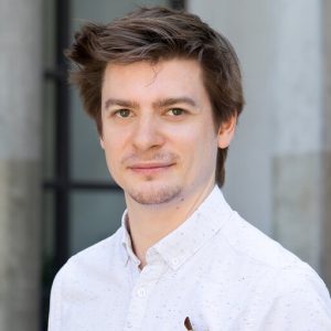
Florent Waltz
University of Basel
Open access to cutting-edge electron and light microscopy
We provide researchers from Europe and beyond with a synergistic portfolio of imaging services including cryo-EM, super-resolution and intravital microscopy to enable new ground-breaking research that crosses the scales of biology.
EMBL Imaging Centre in collaboration with Heidelberg University is offering a 4-day summer school diving into basics of focused ion beam – scanning electron microscopy methods under cryogenic (cryo-FIB-SEM), which allows to study biological samples in their native environment at high-resolution.
Content of the course:
• Theoretical background and practicals delivered by experts in the field
• Sample preparation, lamella preparation and cryo-electron tomography
• Open equipment day to explore further
External invited speaker:

University of Basel
Organisers:

Mattei Team
Edit
Mattei Team
Edit
BioQuant – Heidelberg University
Contact: Anna Steyer anna.steyer@embl.de
To participate to the summer school please register by clicking on the following button. Participation is limited to 9 participants. Deadline for registration is April 1st 2024.
Selected participants will be announced on April 8th 2024.
Sponsor:

Lectures:
Practicals:
Practical 1: Sample preparation (Plunge freezing and high-pressure freezing)
High-resolution electron microscopy requires well vitrified specimen. Depending on the thickness of the sample this can be either accomplished by plunge freezing for thinner specimen, up to a few µm or high-pressure freezing up to ~200 µm.
Transmission electron microscopy (TEM) grids are plasma cleaned to make them less hydrophobic, allowing an even spread of sample. Two different plunge freezers allowing one-side and two-sided blotting used for cells or purified proteins are used to vitrify cells/protein suspensions. A high-pressure freezer is used to freeze thicker specimen e.g. C elegans.
The participants will have the opportunity to use:
Practical 2: Lamella preparation: Serial-FIB/AutoTEM/iFLM
After sample vitrification the TEM grid is introduced into the FIB-SEM and the region of interest is identified. Different milling steps with decreasing beam currents are applied to gradually remove cell material (starting from 1 nA down to 30 pA). Understanding the geometry and cutting approaches while practicing on a real sample will give the best understanding of the challenges, throughput and the possibilities imposed by this method.
The participants will have the opportunity to use:
Practical 3: Cryo-Tomography setup
The TEM grid is introduced into the transmission electron microscope and an overview image of the full grid is taken. After identifying the regions with the lamellae, medium magnification montages are taken to acquire images of the lamella being able to identify and target regions of interest for high-resolution tilt series.
The participants will have the opportunity to use:
Practical 4: Open equipment practical
This practical will allow everybody to spend some more time practice operating an instrument of their choice.
The participants will have the opportunity to use all the equipment used during duration of the course.
| Time (Europe/Berlin) | Speaker | Location |
|---|---|---|
| 9:00-9:15 | Welcome and Introduction Simone Mattei – EMBL | EMBL Imaging Centre room 15-104 |
| 9:15-10:00 | Round table introductions | EMBL Imaging Centre room 15-104 |
| 10:00-10:15 | Q&A | EMBL Imaging Centre room 15-104 |
| 10:15-10:45 | Coffee Break | |
| 10:45-11:30 | Breaking the ice: Basic principals of the FIB-SEM in cryo Anna Steyer – EMBL | EMBL Imaging Centre room 15-104 |
| 11:30-11:45 | Q&A | EMBL Imaging Centre room 15-104 |
| 11:45-13:00 | Lunch break | EMBL Canteen |
| 13:15-18:15 | Practical on Sample preparation plunge freezing and high-pressure freezing (sample preparation rotations of 90′) | EMBL Imaging Centre Labs |
| 19:00 | Dinner | Restaurant Kulturbrauerei |
| Time (Europe/Berlin) | Speaker | Location |
|---|---|---|
| 9:00-9:15 | Feedback discussion and exchanges on practicals | EMBL Imaging Centre room 15-104 |
| 9:15-10:00 | Plunging vs. high-pressure: How to prepare your FIB-SEM sample? Georg Wolff – EMBL | EMBL Imaging Centre room 15-104 |
| 10:00-10:15 | Q&A | EMBL Imaging Centre room 15-104 |
| 10:15-10:45 | Coffee Break | |
| 10:45-11:30 | Cryo-CLEM: Structural analysis of virus -infected cells. Petr Chlanda – Heidelberg University | EMBL Imaging Centre room 15-104 |
| 11:30-11:45 | Q&A | EMBL Imaging Centre room 15-104 |
| 11:45-13:00 | Lunch break | EMBL Canteen |
| 13:15-18:15 | Practical on lamella preparation with a cryo-FIB instrument (lamella preparation rotations of 90′) | EMBL Imaging Centre Labs |
| 19:00 | Free evening |
| Time (Europe/Berlin) | Speaker | Location |
|---|---|---|
| 9:00-9:15 | Feedback discussion and exchanges on practicals | EMBL Imaging Centre room 15-104 |
| 9:15-10:00 | Cryo-Tomography: Tools and concepts in cryo electron tomography. Zhengyi Yang – EMBL | EMBL Imaging Centre room 15-104 |
| 10:00-10:15 | Q & A | EMBL Imaging Centre room 15-104 |
| 10:15-10:45 | Coffee break | |
| 10:45-11:30 | Exploring the architecture of the cell at high-resolution. Florent Waltz – University of Basel | EMBL Imaging Centre room 15-104 |
| 11:30-11:45 | Q & A | EMBL Imaging Centre room 15-104 |
| 11:45-13:00 | Lunch break | EMBL Canteen |
| 13:15-18:15 | Practical cryo electron tomography data collection (cryo-tomography setup) | EMBL Imaging Centre Labs |
| 19:00 | Free evening |
| Time (Europe/Berlin) | Speaker | Location |
|---|---|---|
| 9:00-9:15 | Feedback discussion and exchanges on practicals | EMBL Imaging Centre room 15-104 |
| 9:15-10:15 | Pratical Open Equipment – free Pratical slot | EMBL Imaging Centre Labs |
| 10:15-10:45 | Coffee break | |
| 10:45-11:45 | Pratical Open Equipment – free Pratical slot | EMBL Imaging Centre Labs |
| 11:45-13:00 | Lunch break | EMBL Canteen |
| 13:15-14:45 | Feedback and Closing remarks | EMBL Imaging Centre room 15-104 |
The course is limited to 9 participants. For selection purposes, please note that your application will not be considered without CV and a letter of motivation, stating your background and how this course would be beneficial for your future work.
Selected participants will be announced on April 8th 2024.
There is no registration fee.
Contact: Anna Steyer anna.steyer@embl.de