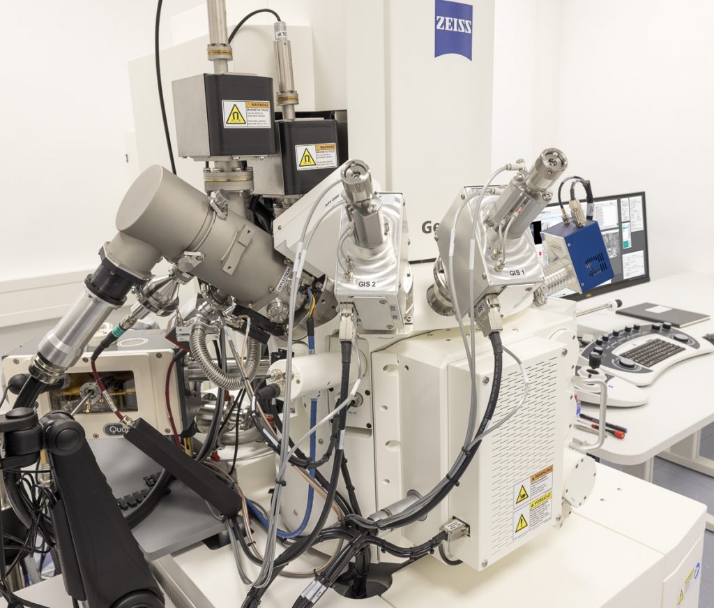
Crossbeam 550
The Crossbeam 550 (focused ion beam-scanning electron microscope) from ZEISS allows data acquisition in 3 dimensions at isotropic nanometer voxel size. Samples from cells to tissues and organisms can be looked at with electron microscopic detail. Chemically fixed, high-pressure frozen and freeze-substituted as well as purely frozen samples can be analyzed. On top of data production, the Crossbeam550 is also equipped to generate cryo-lamellae to be analysed further in a transmission electron microscope e.g. using tomography.
Features
- Isotropic volume acquisition down to a few nm
- Large 3D volume acquisition
- Cryo lamella preparation
Specifications
- Detectors: InLens, EsB and Se
- Quorum Cryo-SEM preparation system
- ZEISS Atlas 5 software
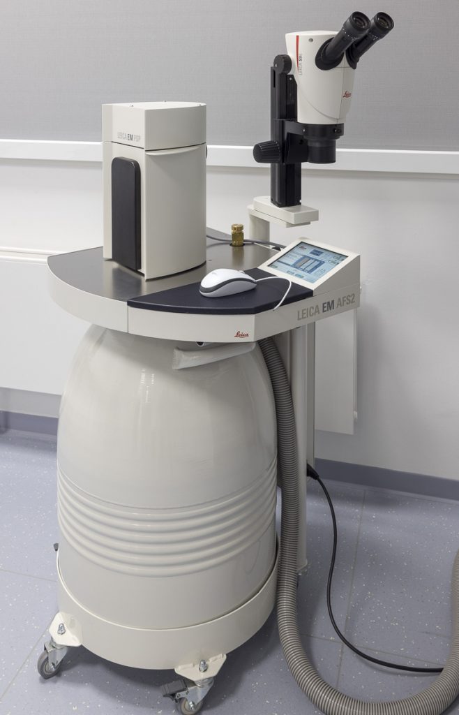
EM AFS2
The EM AFS2 from Leica Microsystems performs freeze substitution and progressive lowering of the temperature (PLT) techniques and allows low temperature embedding and polymerisation of resins.
Features
- Automatic dilution of reagents from stock containers
- Fast UV polymerisation with LED UV lamp for one-step preparation
- Adjustable transfer function of gaseous LN2 to minimise contamination of the specimen
Specifications
- EM FSP (freeze substitution processor), its automatic reagent handling system
EM AFS2 is part of the Coral Life live cell CLEM workflow consisting additionally of the THUNDER Imager Live Cell, and the EM ICE high pressure freezer.
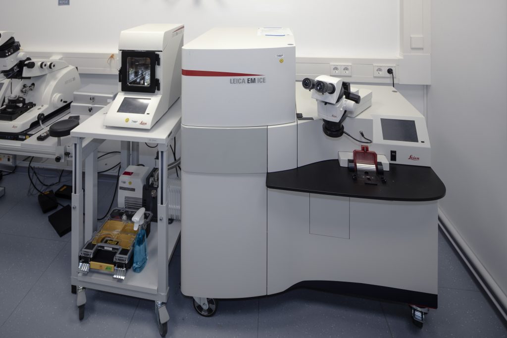
EM ICE
The EM ICE from Leica Microsystems is a modular high-pressure freezing system that can be expanded to enable optical and light stimulation as well as a full live cell CLEM workflow. The freezing principle of the EM-ICE only uses LN2 as cooling liquid, thus preventing alcohol residue in the chamber, on the carrier or the specimens.
Features
- Alcohol-free freezing principle
- From fresh to frozen specimen in 1 second
- Adaptable for live cell imaging. Small modifications in the cartridge preparation area allow fast transfer of specimen between the light microscope and the high-pressure freezer increasing the accuracy of correlation of cellular events in space and time
Specifications
- Electrical and light stimulation module
- Automatic draining of the LN2 Dewar
EM ICE is part of the Coral Life live cell CLEM workflow consisting additionally of the THUNDER Imager Live Cell, and the EM AFS2 for automatic freeze substitution.
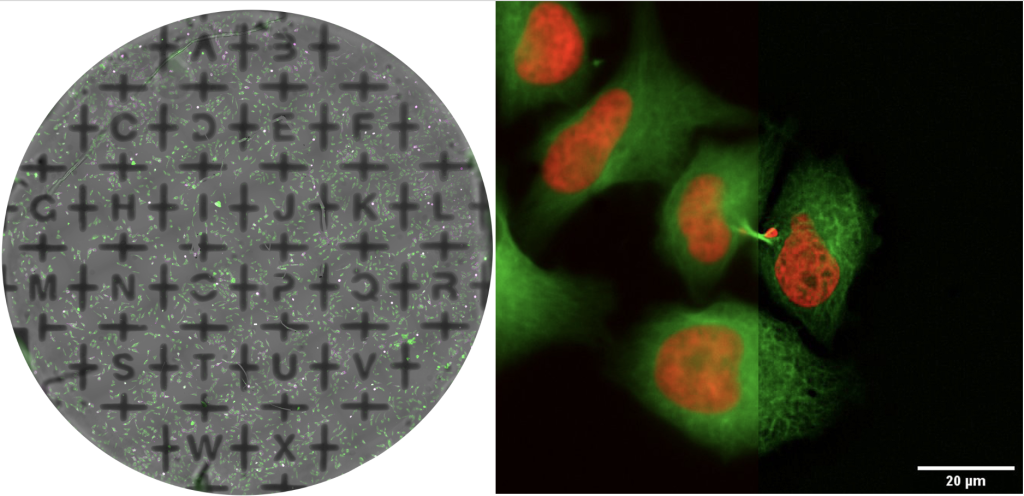
Left: Overview of a saphhire disc mounted in a Samplink chamber. T47D cells labelled for mitochondria (green) and actin filaments (magenta) and brightfield channel representing the carbon masking. Right: High magnification image HeLa Kyoto cells labelled for DNA (red) and microtubules (green) acquired with HC PL APO SAPPHIRE 40x/1,1 W motCORR CS2 objective. Credit: Andreia Pinto, Martin Fritsch / Leica Microsystems
