Edit

EMBL Imaging Centre

Open access to cutting-edge electron and light microscopy

We provide researchers from Europe and beyond with a synergistic portfolio of imaging services including cryo-EM, super-resolution and intravital microscopy to enable new ground-breaking research that crosses the scales of biology.

Super-resolution imaging

Services offered

MINFLUX

Minimal Photon Fluxes Microscopy

SMLM

Single Molecule Localisation Microscopy
STED

Stimulated Emission Depletion Microscopy

Learn more about MINFLUX

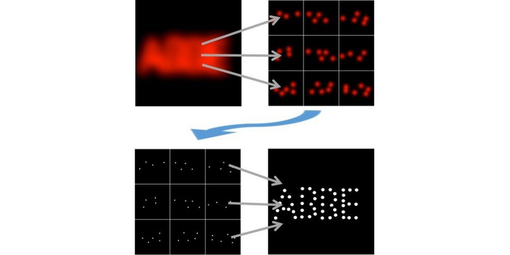
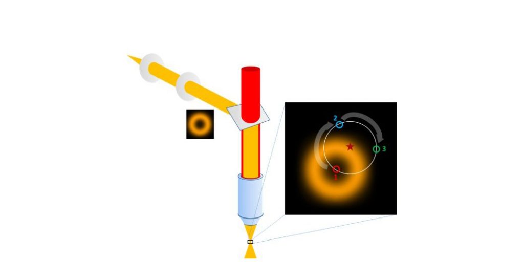
The imaging of minimal photon fluxes combines a coordinate-targeted single point scanning approach with the localization of stochastically activated single fluorophores to provide the currently highest resolving form of super-resolution microscopy.

Upon encountering a single point source signal, the exact position of a single fluorophore is interpolated through the photons collected at different positions of a series of patterns with decreasing search ranges that are executed by the probing beam (a concentric illumination ring with a central minimum) to home in on the fluorophore position. The combination of beam targeting with single molecule detection thus allows a localization accuracy in the nanometer range while only requiring a minimal number of photon detections overall. The MINFLUX principle can be applied for highly resolved imaging (stochastically activated fluorophores in a sample are imaged sequentially and integrated into a set of coordinates) as well as for the dynamic tracing of a single fluorescent molecule at unprecedented temporal and spatial resolution.

Learn more about SMLM

In SMLM is based on the fact that in diffraction-limited optics the position of a single point (i.e. a single molecule) can be determined with much higher accuracy than the minimal resolvable distance between two points.

Applying a fit for the central maximum of a single point source can thus determine its position with an accuracy that can be an order of magnitude higher than the optical resolution of the imaging system used. This cannot be applied in standard imaging as single point sources cannot be separated from their neighbours while all of them are detected together. In SMLM the label is made sufficiently scarce for the detection of single molecule positions by either the sparse photoconversion of single molecules to an active state (Photoactivation Localisation Microscopy, PALM), by the induction of transient darkstates in a vast majority of the fluorophores in the image field with only randomly distributed single emitters on at any time (Stochastic Optical Reconstruction Microscopy, STORM) or by the transient binding of diluted fluorescent tags to their targets (Points Accumulation for Imaging in Nanoscale Topography, PAINT).

Learn more about STED

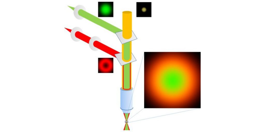
In STED microscopy, an excited fluorescent molecule is induced to return to the ground state through the interaction with a photon of a wavelength inside the fluorophore’s emission spectrum.

This process is called stimulated emission. The combination of a focused excitation beam with a phase-shifted depletion beam in the same position creates a central excitation volume that is surrounded by a concentric ring of depletion light that deactivates fluorophores in its area and thus will reduce the effective fluorescence detection volume. In combination with confocal scanning this allows imaging below the diffraction limit. The resolution improvement is determined by the intensity of the applied depletion and can either be applied directly or be further enhanced by analysing the distribution of shortened fluorescence lifetimes that are caused by the interaction with the depletion beam.

MINFLUX

MINFLUX from Abberior Instruments enables imaging of a broad range of biological samples with a spatial resolution in the single nanometer range. This allows to study the structure of large multimolecular protein complexes, such as nuclear pores with unprecedented 3D resolution. 

Additionally, MINFLUX tracking allows to study movement of single molecules in living cells with up to 10 kHz sampling rate (one localisation each 100 µs, which is up to 100 x faster than conventional camera-based tracking). 


Features:

  • MINFLUX imaging with 1-3 nm localisation precision in 2D/3D
  • Tracking of single molecules with up to 10 kHz featuring <20 nm resolution 
  • Nanometre-level active sample stabilization system based on fiducial markers in the sample

 Specifications:

  • Laser lines: MINFLUX: 640 nm, 561 nm, Confocal: 488 nm, 405 nm
  • Two-colour ratiometric MINFLUX imaging with 640 nm or 560 nm excitation
  • IX83 inverted microscope stand equipped with a 1.4 NA 100x oil objective


Credit: Sebastian Schnorrenberg/EMBL
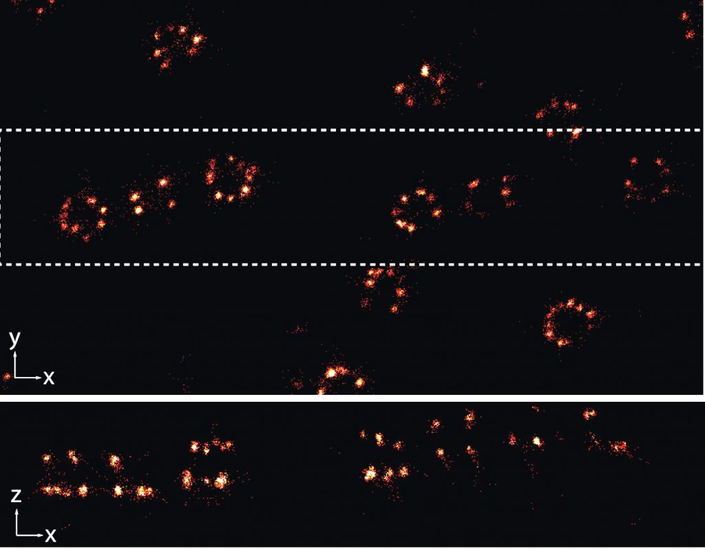
U2OS cells: 3D MINFLUX dataset of nuclear pore complex protein Nup96 with Alexa647 labelled SNAP-tag. Shown is the lateral (xy) view of the sample and a sideways (xz) projection of the highlighted subregion. Credit: Sebastian Schnorrenberg/EMBL
Credit: Stuart Ingham/EMBL.
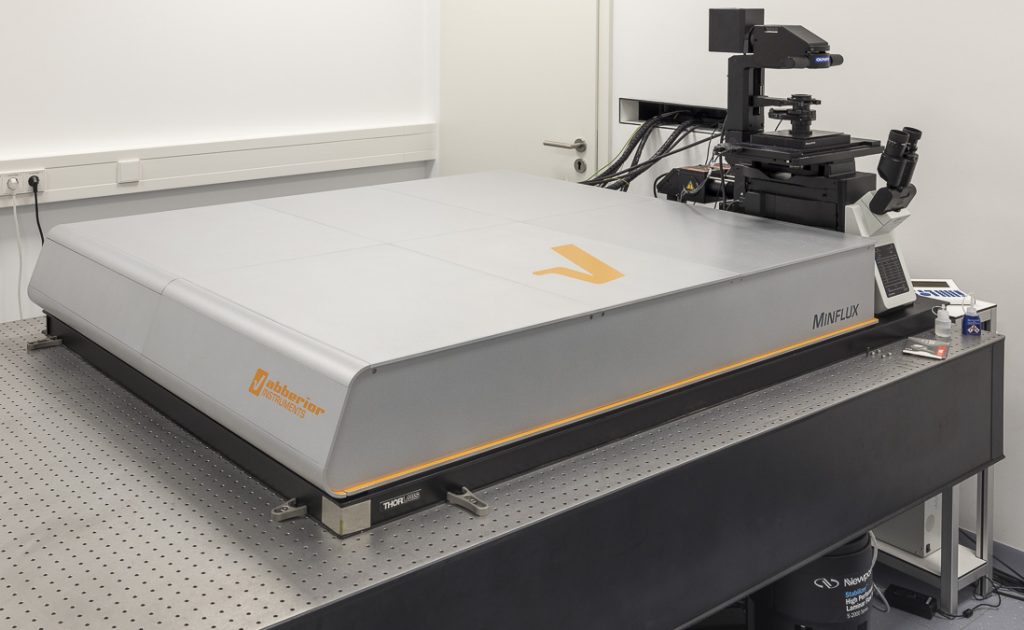
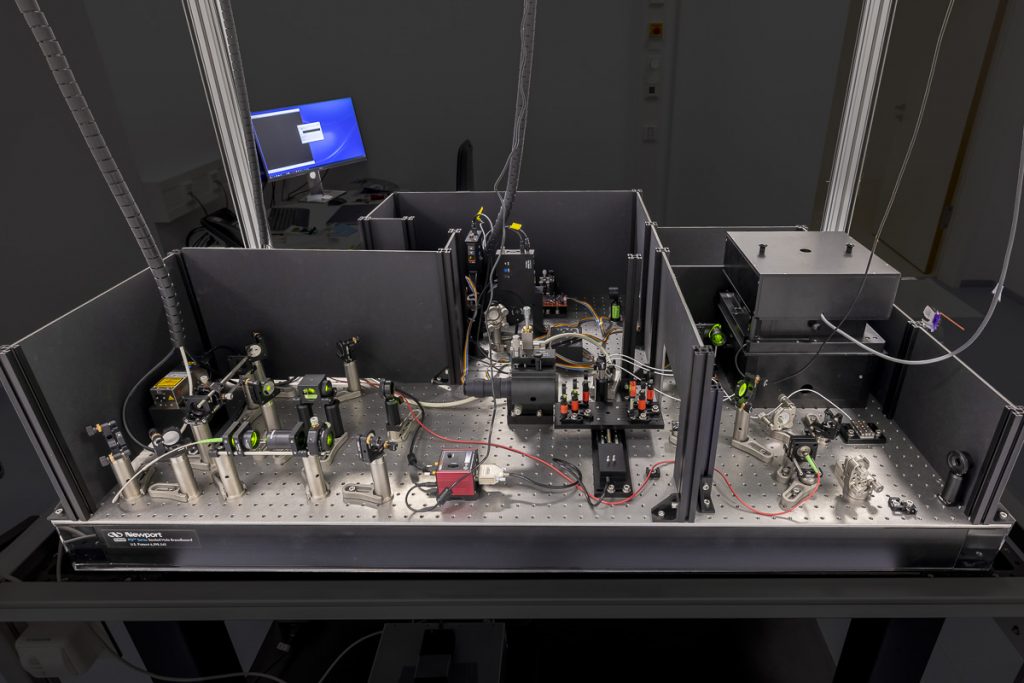
Abberior MINFLUX. Credit: Stuart Bailey/EMBL

3D-SMLM

Three-dimensional single-molecule localisation microscopy (3D-SMLM) is an advanced light microscopy methodology based on localisation of sparse single-molecule emitters for computational reconstruction of a super-resolved image. The 3D-SMLM at the EMBL IC has been developed in collaboration with the lab of Jonas Ries at EMBL. At the core of the microscope is an extremely stable inverted microscope. (f)PALM/(d)STORM workflows can be accommodated and additional functionalities will be added dependent on user need (e.g. support for live-cell imaging/PAINT/SOFI and others).


Features

  • Ratiometric multi-colour imaging (e.g. simultaneous multi-colour imaging of spectrally overlapping fluorophores)
  • Homogenised Epi-/HILO/TIRF illumination with variable illuminated field ca. ⌀20 – 70 microns, Super-resolution over large field of views (up to ⌀70 microns)
  • Option for 3D imaging via astigmatism
  • < 10 nm localisation precision (in x,y, dependent on fluorophore, imaging mode, < 20 nm in z)
  • Automated high-throughput imaging (e.g. multiple field of views)

Specifications

  • 4 lasers generating ca. 100 mW at source (Toptica iChrome MLE, 405, 488, 561, 640 nm), additional booster laser at 640 nm (Toptica iBeam Smart 200 mW).
  • Back-thinned sCMOS camera (Hamamatsu Orca Fusion BT >95% QE, <0.7 e- read noise)
  • Focus locking via reflection of NIR laser (Toptica iBeam Smart, 785 nm)
  • Motorized XY nanopositioning stage (SmarAct), Piezo actuator (PI PIFOC, P-726) for objective.
  • Olympus 100x/1.45 (oil), 100x/1.35 (silicone oil) objective lenses
  • The microscope can be configured for the needs of the experiment via a range of switchable optical elements. 3D imaging is achieved using the astigmatic method and analysis/reconstruction is achieved using the SMAP toolset
Credit: Merle Hantsche-Grininger/EMBL
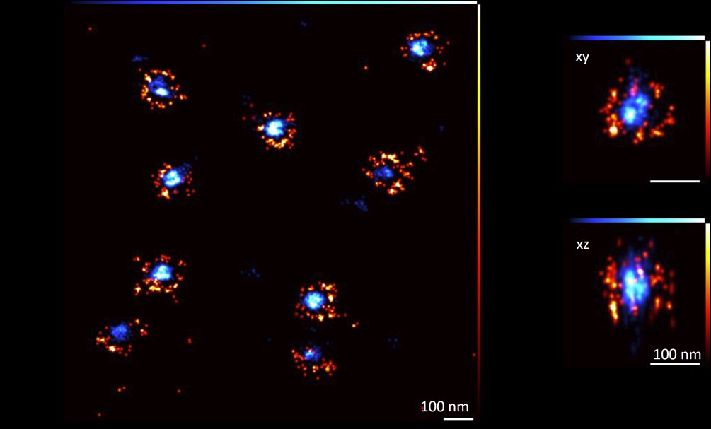
3D-SMLM images of U2OS Nup96-mEGFP cells stained with Q nanobody AF647 (orange) and WGA-CF680 (blue). Left: Zoom in on several nuclear pores. Top right: an XY view of a single nuclear pore showing the ring structure and separation of the two dyes. Bottom right: an XZ view of the same nuclear pore, showing
that the two stacked rings can be resolved. Scale bars, 100 nm. Credit: Merle Hantsche-Grininger/EMBL.
Credit: Stuart Ingham/EMBL.
3D-SMLM at IC. Credit: Stuart Ingham/EMBL.

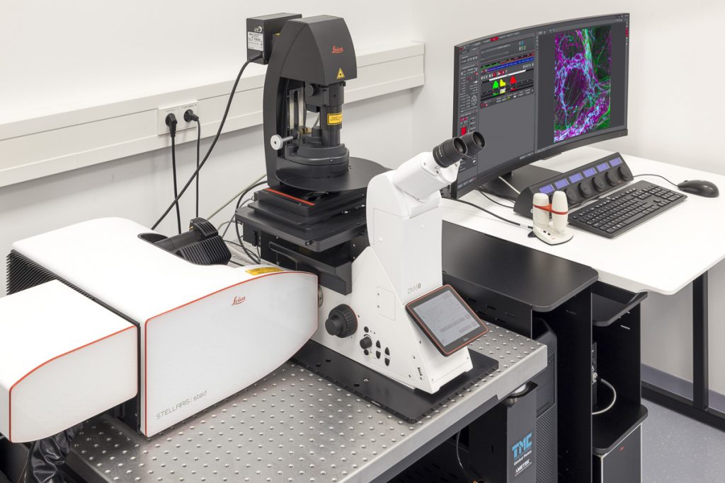
STELLARIS 8 STED Falcon

STELLARIS 8 STED with TauSTED from Leica Microsystems allows the study of multiple dynamic events simultaneously and investigation of molecular relationships and mechanisms within the cellular context.

FALCON (FAst Lifetime CONtrast) includes phasors for quantitative FLIM analysis. It is a fully integrated solution for fluorescence lifetime imaging (FLIM) and enables video-rate image acquisition for rapid kinetic studies in live cells. 


Features

  • Resolving molecular relationships in specimens
  • Multiple simultaneous events can be studied at the nanoscale
  • 2D and 3D STED imaging
  • Significantly reduced light doses due to TauSTED
  • Advanced STED-FLIM options with STED and FALCON (full FLIM quantitative tools, phasor FLIM, species separation based on phasor analysis)
  • Advanced STED-FCS options
  • Fast validation of results on a single platform, from confocal to super-resolution LIGHTNING and STED
  • With FALCON it is possible to:
    • follow fast molecular interactions via FLIM-FRET (Förster resonance energy transfer).
    • use biosensors to detect microenvironmental changes, such as pH or ion concentration.
    • apply lifetime contrast to separate multiple fluorophores.
    • do analysis using phasor FLIM provides a 2D visualisation of lifetime components. With FLIM phasors you can follow microenvironmental changes, select components to multiplex signal (phasor separation), and determine FRET efficiency.

Specifications

  • Five spectrally tunable Power HyD sensitive photon counting detectors (2 HyD S, 2 HyD X, 1 HyD R)
  • Confocal: tunable White Light Lasers (WLL) 440-790 nm, 405 nm STED depletion: 592 nm, 660 nm, 775 nm
  • 8kHz Resonant Scanner
  • Total system dead-time: 1.5 ns
  • TauSTED: Tunable resolution based on lifetime (depending on sample and fluorophore: <30 nm (lateral) and <100 nm (axial). Automatic lifetime-based background suppression algorithm. Light dose reduction (WLL excitation) for all STED lines (592, 660, 775 nm). Available for 2D and 3D STED in live and in fixed specimens, also for multicolor applications. Automated workflow integrated in the LAS X software.
  • The STED WHITE glycerol and water objective lenses with motCORR technology provide adaptive optical correction for aberrations introduced by sample inhomogeneities and refractive-index mismatch. The STED WHITE objective lenses provide a working distance of 300 µm:
    • HC PL APO 86x/1.20 W motCORR STED WHITE
    • HC PL APO 93x/1.30 GLYC motCORR STED WHITE
    • HC PL APO 100x/1.40 OIL STED WHITE
Credit: Timo Zimmermann/EMBL.
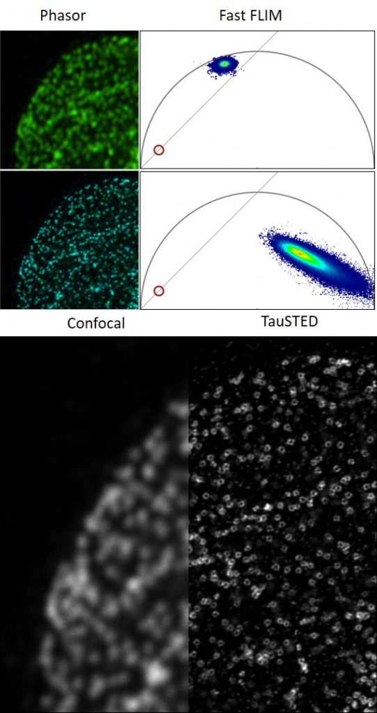
U2OS cells: FLIM images under confocal and STED conditions of nuclear pore protein Nup96 and their corresponding phasor plots of lifetime information. TauSTED image of the same cell. Images taken with Leica Stellaris 8 STED Falcon. Credit: Timo Zimmermann/EMBL.
Credit: Stuart Ingham/EMBL.
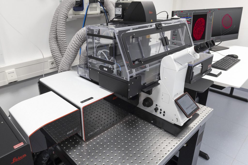
Leica STELLARIS 8 STED Falcon. Credit: Stuart Ingham/EMBL.

STELLARIS STED

STELLARIS STED equipped with TauSTED from Leica Microsystems allows the study of multiple dynamic events simultaneously and investigation of molecular relationships and mechanisms within the cellular context.


Features

  • Resolving molecular relationships in specimens
  • Multiple simultaneous events can be studied at the nanoscale
  • 2D and 3D STED imaging
  • Significantly reduced light doses due to TauSTED
  • Fast validation of results on a single platform, from confocal to super-resolution LIGHTNING and STED

Specifications

  • Five spectrally tunable Power HyD sensitive photon counting detectors (2 HyD S, 2 HyD X, 1 HyD R)
  • Confocal: tunable White Light Lasers (WLL) 440-790 nm, 405 nm STED depletion: 592 nm, 660 nm, 775 nm
  • 8kHz Resonant Scanner
  • Total system dead-time: 1.5 ns
  • TauSTED: Tunable resolution based on lifetime (depending on sample and fluorophore: <30 nm (lateral) and <100 nm (axial). Automatic lifetime-based background suppression algorithm. Light dose reduction (WLL excitation) for all STED lines (592, 660, 775 nm). Available for 2D and 3D STED in live and in fixed specimens, also for multicolor applications. Automated workflow integrated in the LAS X software.
  • The STED WHITE glycerol and water objective lenses with motCORR technology provide adaptive optical correction for aberrations introduced by sample inhomogeneities and refractive-index mismatch. The STED WHITE objective lenses provide a working distance of 300 µm:
    • HC PL APO 86x/1.20 W motCORR STED WHITE
    • HC PL APO 93x/1.30 GLYC motCORR STED WHITE
    • HC PL APO 100x/1.40 OIL STED WHITE
Credit: Timo Zimmermann/EMBL
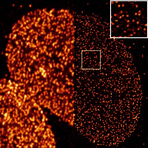
U2OS cells: Comparison of confocal and TauSTED images of nuclear pore protein Nup96. Images taken with Leica Stellaris STED. Credit: Timo Zimmermann/EMBL.
Credit: Stuart Ingham/EMBL.
Leica STELLARIS STED. Credit: Stuart Ingham/EMBL.
Edit