Calina Glynn
Rosalind Franklin Institute
Open access to cutting-edge electron and light microscopy
We provide researchers from Europe and beyond with a synergistic portfolio of imaging services including cryo-EM, super-resolution and intravital microscopy to enable new ground-breaking research that crosses the scales of biology.
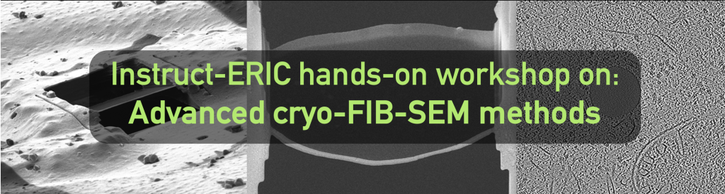
EMBL Imaging Centre is offering a 5-day course (21-25 July 2025) diving into advanced operations of focused ion beam – scanning electron microscopy methods under cryogenic conditions (cryo-FIB-SEM), which allows to study biological samples in their native environment at high-resolution.
Content of the course:
• Theoretical background and practicals delivered by experts in the field
• Advanced sample preparation using waffle method, advanced lamella preparation, correlative workflows and volume-imaging
• Open equipment day to explore further
External invited speaker:
Rosalind Franklin Institute
Organisers:
Contact:
Anna Steyer (anna.steyer@embl.de)
To participate in this course please register by clicking on the following button. The course is limited to 12 participants. Deadline for registration is 12th May 2025. Selected participants will be announced on 19th May 2025. For selection purposes, please note that your application will not be considered without CV and a letter of motivation, stating your background and how this course would be beneficial for your future work.
There is no registration fee. Selected participants from an Instruct member state will receive 550€ travel and accommodation grant.
Funded by:
Practical 1: Sample preparation by Waffle method (HPF and cryo-LM)
Standard cellular lamella production by cryo-FIB milling poses major challenges when preparing small, unicellular samples (e.g. bacteria or protists) that often result too thin to offer proper lateral support to the generated lamella. At the same time, thick multi-cellular specimen (e.g. 3D organoids, C. elegans) cannot be properly preserved by simple plunge-freezing preparation, where the limited heat transfer results in crystalline ice formation. Both challenges have been recently overcome by introducing a new method, termed “waffle method” (Kelley et al. 2022, Nature), allowing on-grid lamella preparation after high-pressure freezing without the need for lift-out.
The waffle method starts with the preparation of the high-pressure freezing (HPF) carrier and glow discharging of the transmission electron microscopy (TEM) grids. For the actual freezing by high-pressure freezing, a sandwich of HPF carriers surrounding a TEM grid where the sample was applied is assembled and frozen. After samples vitrification, the TEM grid is gently separated from the carrier and can now be checked by cryo-LM. Different objectives (5x, 10x and 100x) in combination with reflected light and fluorescence microscopy will reveal the structure of interest and its location on the grid.
The participants will have the opportunity to use:
Practical 2: Specifics of waffle samples in the FIB-SEM (cryo-lamella preparation)
After sample vitrification the TEM grid is introduced into the FIB-SEM and the region of interest is identified. Different milling steps with decreasing beam currents are applied from different angles to remove material (starting from 15 nA down to 30 pA). Understanding the geometry and cutting approaches while practicing on a real sample will give the best understanding of the challenges, throughput and the possibilities imposed by this method.
The participants will have the opportunity to use:
Practical 3: Automating lamella preparation and targeting using Serial-FIB
Vitrified cells on a TEM grid including fiducials will be introduced into the FIB-SEM to target regions of interest in 3D (3DCT toolbox). Multiple positions will be set-up to be milled automatically with the focused ion beam to get lamellae suitable for tomographic data acquisition.
The participants will have the opportunity to use:
Practical 4: Cryo-volume imaging using FIB-SEM
Being able to visualize fully hydrated samples as close as possible to native conditions with volume microscopy has seen an increasing interest by the community. Samples that cannot be prepared well for traditional electron microscopy due to their intrinsic properties (e.g. biominerals or biofilms) greatly benefit from this imaging method. The combination of cryo volume imaging followed by lamella preparation can also be used to target structures of interests.
Samples will be introduced into the FIB-SEM and prepared for volume data acquisition, including sputtering, opening the sample surface exposing a cross-section and setting up a volume data acquisition. Milling and imaging will be alternated to acquire a dataset through the region of interest under native conditions.
The participants will have the opportunity to use: