LSM900 Airyscan2
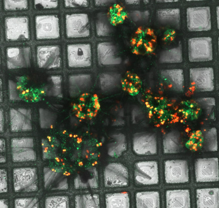
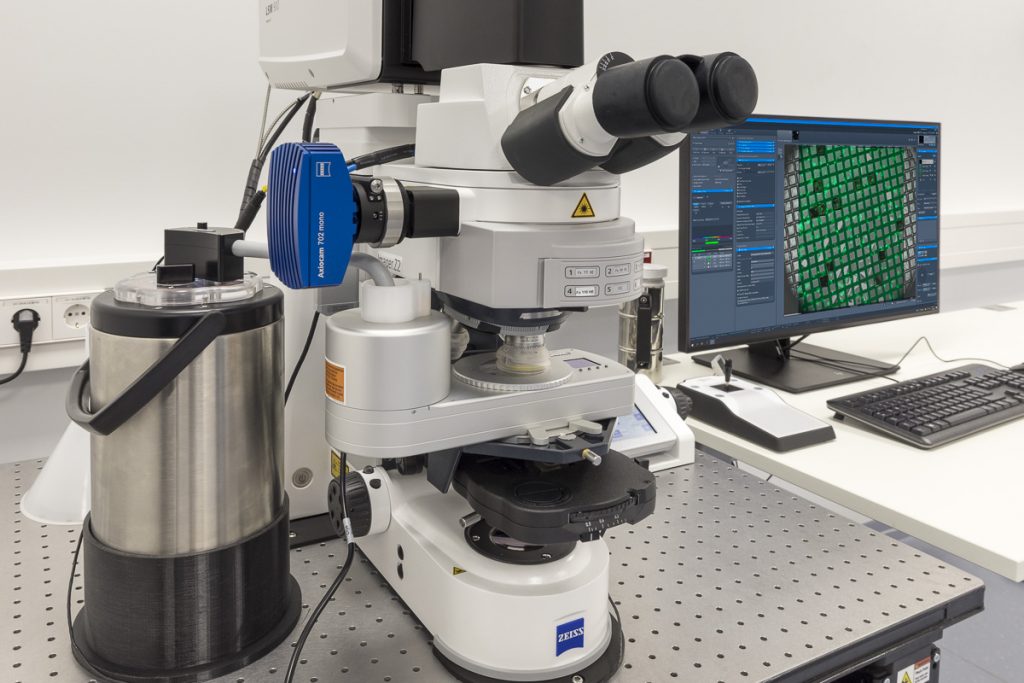
The LSM900 Airyscan2 from ZEISS is equipped with an Linkam cryo stage and configured for high-resolution confocal imaging of fluorescent sample. The Airyscan module enables super-resolution imaging without introducing high laser power or devitrifying the sample under liquid nitrogen temperature. In combination with the cryo Linkam stage, it is able to accommodate frozen samples on plunge-frozen EM grids or high-pressure frozen planchettes. The sample holder that fits both, the Linkam stage as well as the ZEISS Crossbeam enables the correlation between fluorescent light and scanning electron microscope.
Features
- Imaging frozen sample under cryogenic temperature
- Full grid montage tiling
- Triple / quadruple filter sets enables multi-channel imaging simultaneously
- Airyscan super-resolution imaging
- Correlative workflow from light to electron microscope
Specifications
- Calibri 7 LED source that covers 7 fluorescent channels (385, 430, 475, 555, 590, 630 and 735nm)
- 4 laser sources (405, 488, 561 and 641nm)
- Filter sets 90HE, 92HE, 110HE and 77HE
- Airyscan calibrated 100x/0.75 air objective to achieve 250nm lateral resolution


STELLARIS 5 cryo
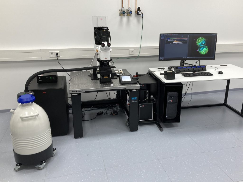
STELLARIS 5 Cryo confocal light microscope from Leica Microsystems offers to image vitrified (e.g., plunge frozen or high pressure frozen) fluorescent samples under constant cryogenic conditions. The STELLARIS 5 Cryo imaging platform may offer variable excitation and detection possibilities not only to define matching imaging conditions, but also to determine specific signals in a cryogenic environment.
Features
- TauSense technology on the STELLARIS cryo imaging platform gives access to specific fluorescence photon arrival-time information, such as reflection of grids, fluorophore arrival-time and autofluorescence arrival-time in cryogenic conditions. It helps to improve specific signal detection, removing unwanted background or grid reflection and it expands the combination of fluorescent signals beyond the spectral options.
- Image quality beyond the visible detection range in real time and at full speed in combination with the enhanced spatial resolution using LIGHTNING super-resolution module.
- Based on an upright microscopy stand dedicated for cryo microscopy imaging, but also for room temperature imaging and dip-in applications.
Specifications
- STELLARIS 5 Scanhead with 3 HyD S detectors
- Laser supply unit with super-continuum laser (485 – 685 nm), diode laser lines: 405 nm, 448 nm, 488 nm, 561 nm, 638 nm.
- K5 sCMOS camera
- Upright DM6 confocal fixed stage microscope
- HC PL APO 50x/0,90 CRYO CLEM cryo objective lens
- Dipping objective lens for upright room temperature applications
- Märzhäuser stage equipped with Cryo stage or sample holder for RT imaging
- 25 l dewar with cryo pump and cryo control unit
STELLARIS 5 Cryo is part of the Coral Cryo CLEM workflow consisting additionally of EM GP2, EM VCM plus EM VCT500 Cryo transfer shuttle or EM ACE600 coater plus EM VCT 500 cryo transfer shuttle.


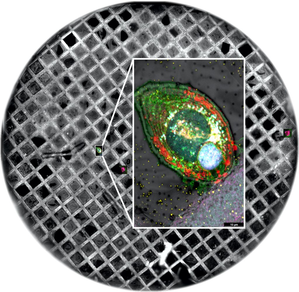
Budding yeast Saccharomyces cerevisiae stained against nuclei and actin imaged with the Coral Cryo CLEM workflow. From left to right: combination of SEM and confocal grid overview (left), Lamella and high-resolution confocal and cryo TEM image data (right).
Credit: Andreia Pinto, Martin Fritsch / Leica Microsystems