Yannick Schwab
Team Leader and Head of Electron Microscopy Core Facility
ORCID: 0000-0001-8027-1836
EditThe facility provides advanced expertise in electron microscopy, from sample preparation to image analysis, for a wide variety of biological samples.
Team Leader and Head of Electron Microscopy Core Facility
ORCID: 0000-0001-8027-1836
EditThe EMCF provides advanced expertise in cellular electron microscopy (EM) at room temperature: from sample preparation to image analysis and for a large variety of biological samples ranging from bacterial cells to (small) multicellular organisms. Our main focus is on ultrastructural analysis in 2D and 3D, immuno-EM and correlative multimodal imaging, combining EM with light microscopy (CLEM), and X-ray microscopy (CXEM) or both (CLXEM). Our staff can help you define optimal experimental conditions for your project.
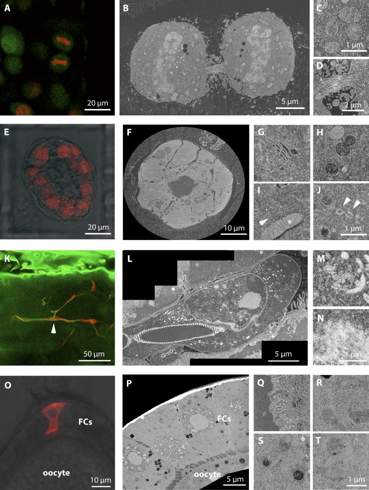
One of our strengths is the on-section CLEM technique, which tracks the signal of fluorescent proteins in resin sections with high precision. We have used this method for a number of studies (Groza et al. 2024, Schreier et al. 2022) and published a detailed step-by-step video tutorial on this method.We are also hosting an international course covering the methodology every second year.
Our facility is also exploiting different volume EM techniques such as serial block face – scanning electron microscopy (SBF-SEM) and focused ion beam – scanning electron microscopy (FIB-SEM), to visualize the 3D ultrastructure of organelles at nanometer resolution across micron scales. For the precise targeting of the ROI in a 3D volume, we are routinely using correlative approaches combining light (see below) and X-ray imaging together with EM. The X-ray imaging is done in the facility with a tabletop system and when going for higher resolution in collaboration with Liz Duke on the EMBL beamline P14 at the PETRA III synchrotron in Hamburg.
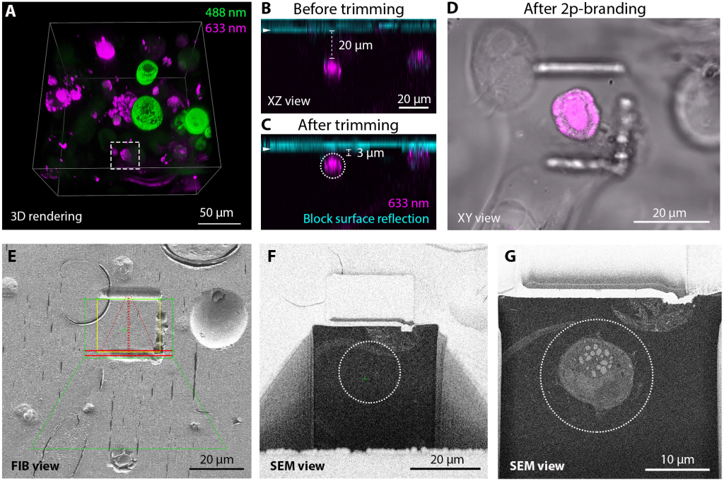
We are also involved in method development, which is heavily connected to our collaborations with groups here at the EMBL. An example of this is the development of a high-precision targeting methods for volume electron microscopy, together with the Ephrussi, Jechlinger, Leptin and Mahamid groups (Ronchi et al., 2021). The strategy relies on fluorescence preservation during sample preparation and targeted trimming guided by confocal maps of the fluorescence signal in the resin block. Laser branding is used to create landmarks on the block surface to position the FIB-SEM acquisition.
Please note that single-particle and tomography cryo-EM for structural biology projects are offered through the cryo-EM service platform and the EMBL IC
.
The small GTPase Ran Defines nuclear pore complex asymmetry
Sachweh J, Börmel M, Klumpe S, Becker A, Taniguchi R, Kubańska MA, Pintschovius V, Kaindl A, Plitzko JM, Wilfling F, Beck M and Hampoelz B. Cell., 2025 Oct 16;188(21):5931-5946.
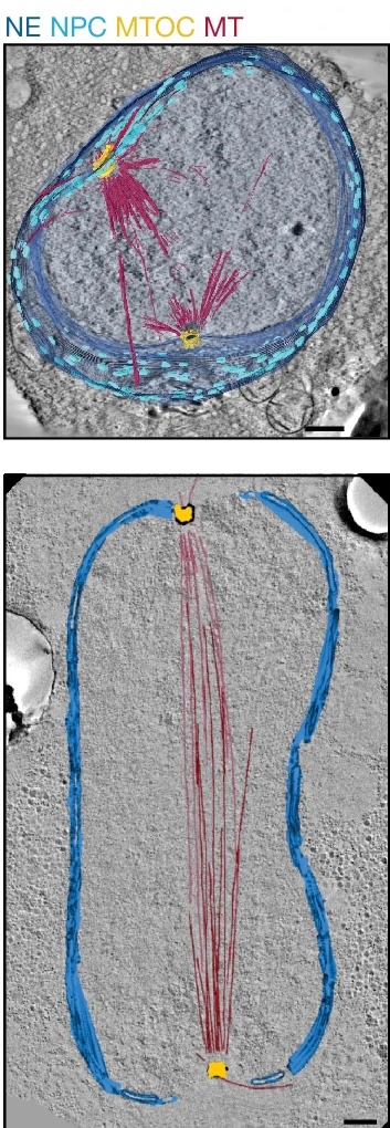
Life-cycle-coupled evolution of mitosis in close relatives of animals
Shah H, Olivetta M, Bhickta C, Ronchi P, Trupinić M, Tromer EC, Tolić IM, Schwab Y, Dudin O and Dey G. Nature., 2024 Jun;630(8015):116-122.
Targeted volume correlative light and electron microscopy of an environmental marine microorganism
Mocaer K, Mizzon G, Gunkel M, Halavatyi A, Steyer A, Oorschot V, Schorb M, Le Kieffre C, Yee DP, Chevalier F, Gallet B, Decelle J, Schwab Y and Ronchi P. J Cell Sci., 2023 Aug 1;136(15):jcs261355.
High-precision targeting workflow for volume electron microscopy.
Ronchi P, Mizzon G, Machado P, D’Imprima E, Best BT, Cassella L, Schnorrenberg S, Montero MG, Jechlinger M, Ephrussi A, Leptin M, Mahamid J, Schwab Y. J Cell Biol. 2021 Sep 6;220(9):e202104069.
If you would like to apply as a visitor in the EMCF, please contact us at emcf@embl.de including a short overview of your project. We’ll discuss the feasibility of your project internally and in case of a positive evaluation we will contact you for a more detailed meeting over the phone, via Zoom or video conference.
The EMCF currently participates in several initiatives supporting visitor access:
The EMCF is part of the EMBL node in Euro-BioImaging, together with the Advanced Light Microscopy Facility (ALMF) at EMBL Heidelberg and the Mesoscopic Imaging Facility (MIF) at EMBL Barcelona. The EMBL node offers open access to a broad range of state-of-the-art technologies in biological imaging for life scientists. Euro-Bioimaging is a platform for user access and a resource for providing different funding opportunities.
The Fellowships support travel and accommodation costs of young scientists visiting EMBL Core Facilities at the different EMBL sites.
Global Bioimaging offers the International Job Shadowing program to technical and managerial staff of imaging core facilities. These visits provide the user with the opportunity to visit an imaging core facility abroad, learn from their peers and provides a fantastic networking opportunity.