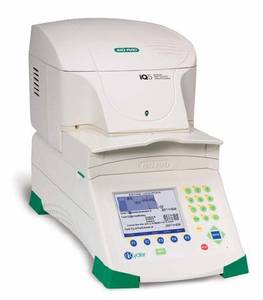
Thermofluor
In this method, the protein is incubated with a dye that only binds hydrophobic protein regions, where it becomes highly fluorescent. The protein is gradually heated, in order to slowly unfold, exposing these hydrophobic patches.
Target proteins used for crystallization or SAXS can be studied by thermal denaturation assays. The melting point of a protein (Tm) is influenced not only by the nature of the protein itself but also by its chemical environment (e.g. pH, salt nature and concentration, additives, presence of natural or synthetic ligands, etc.). It is possible determine optimum conditions stabilising the protein fold.
Two available instruments to perform thermal denaturation assays:

In this method, the protein is incubated with a dye that only binds hydrophobic protein regions, where it becomes highly fluorescent. The protein is gradually heated, in order to slowly unfold, exposing these hydrophobic patches.

NanoDSF is an advanced Differential Scanning Fluorimetry method for measuring ultra-high resolution protein stability using intrinsic tryptophan or tyrosine fluorescence.
The RUBIC buffer screen has been designed at the SPC facility and is now distributed under licence by Molecular Dimensions.
Features of the RUBIC Buffer Screen:
Sample requirement for TF:
Sample requirements for nanoDSF:
Contact SPC to discuss your experiment in detail
The additive screen aims to explore the effect of several additives on the thermal stability of the proteins. We recommend to perform this screen to complement the BUFFER screen.
Applications:
Sample requirement for TF:
Sample requirements for nanoDSF:
Contact SPC to discuss your experiment in detail
Determination of the Molecular Mass of your protein sample.
CovalX HM4 High-Mass System enables:
Protein complex analysis
Intact Protein Analysis (upto 2MDa)
Contact us for detailed protocol, instrument booking, training, pricing, etc.