3D structure of the ribosome
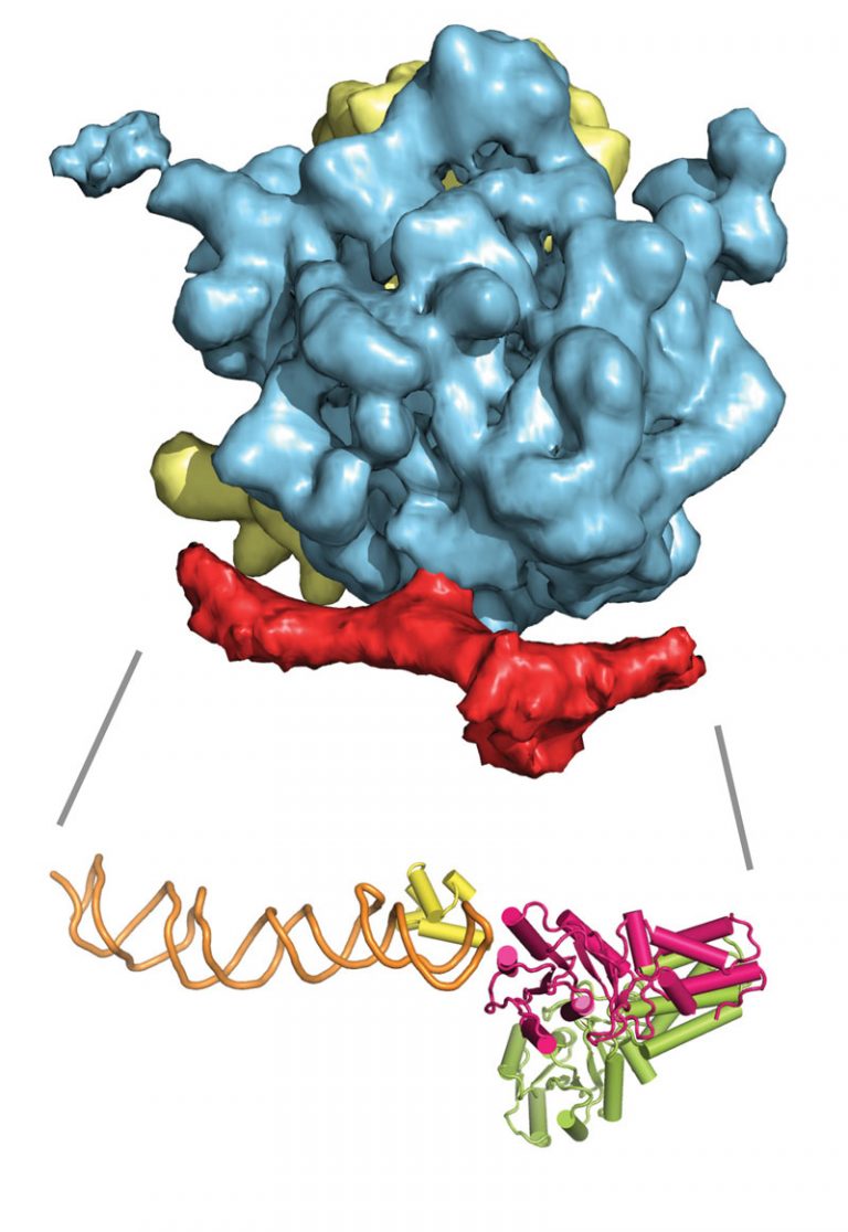
This cryo-electron microscopy image shows the 3D structure of the ribosome (yellow/blue) bound to the signal recognition particle (SRP) and the SRP receptor (both in red). Below it is an atomic model of SRP (green-yellow/orange) and its receptor (pink). Image credits: EMBL/Schaffitzel.
Download:
full (800x1163) | thumbnail (150x150) | medium (206x300) | medium_large (768x1116) | large (704x1024)