Uncovering secrets of life in the ocean
Researchers unravel how the very first eyes in evolution might have worked and how they guide the swimming of marine plankton towards light

The best-selling novel The swarm captured the imagination of countless readers with the fascination of marine life. But it also showed how little we understand life in the depth of the ocean. Scientists at the European Molecular Biology Laboratory (EMBL) and the Max Planck Institute (MPI) for Developmental Biology now explain the remarkable ability of marine zooplankton to swim towards light. Their study, published in the current issue of Nature, reveals how simple eyes of only two cells sense the direction of light and guide movement towards it. The key is a nerve that connects the eyes directly to the cells that mediate swimming. The research also provides new insights into what the first eyes in animal evolution might have looked like and what their function was.
Larvae of marine invertebrates – worms, sponges, jellyfish – have the simplest eyes that exist. They consist of no more than two cells: a photoreceptor cell and a pigment cell. These minimal eyes, called eyespots, resemble the ‘proto-eyes’ suggested by Charles Darwin as the first eyes to appear in animal evolution. They cannot form images but allow the animal to sense the direction of light.This ability is crucial for phototaxis – the swimming towards light exhibited by many zooplankton larvae. Myriads of planktonic animals travel guided by light every day. Their movements drive the biggest transport of biomass on earth.
“For a long time nobody knew how the animals do phototaxis with their simple eyes and nervous system,” explains Detlev Arendt, whose team carried out the research at EMBL. “We assume that the first eyes in the animal kingdom evolved for exactly this purpose. Understanding phototaxis thus unravels the first steps of eye evolution.”
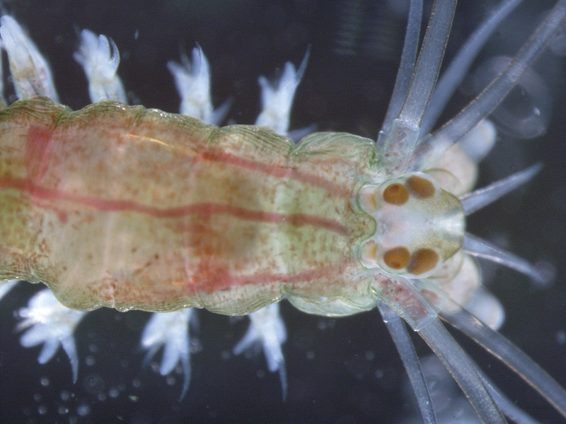
Studying the larvae of the marine ragworm Platynereis dumerilii, the scientists found that a nerve connects the photoreceptor cell of the eyespot and the cells that bring about the swimming motion of the larvae. The photoreceptor detects light and converts it into an electrical signal that travels down its neural projection, which makes a connection with a band of cells endowed with cilia. These cilia – thin, hair-like projections – beat to displace water and bring about movement.
Shining light selectively on one eyespot changes the beating of the adjacent cilia. The resulting local changes in water flow are sufficient to alter the direction of swimming, computer simulations of larval swimming show.
The second eyespot cell, the pigment cell, confers the directional sensitivity to light. It absorbs light and casts a shadow over the photoreceptor. The shape of this shadow varies according to the position of the light source and is communicated to the cilia through the signal of the photoreceptor.
“Platynereis can be considered a living fossil,” says Gáspár Jékely, former member of Arendt’s lab who now heads a group at the MPI for Developmental Biology, “it still lives in the same environment as its ancestors millions of years ago and has preserved many ancestral features. Studying the eyespots of its larva is probably the closest we can get to figuring out what eyes looked like when they first evolved.”
It is likely that the close coupling of light sensor to cilia marks an important, early landmark in the evolution of animal eyes. Many contemporary marine invertebrates still employ the strategy for phototaxis.


