Not a galaxy far, far away

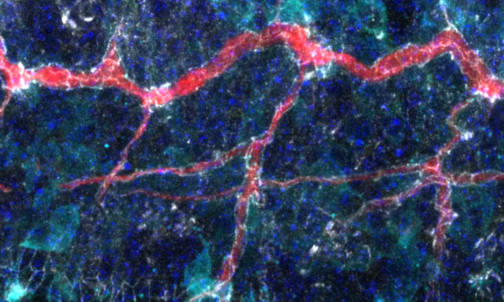
While this may seem like a nebula made up of interstellar clouds of dust and ionised gases, this image isn’t of a galaxy beyond the Milky Way. Rather it captures something quite common on our own planet, visually probing a fruit fly embryo midway through development.
Members of EMBL’s Leptin Group collected, fixed, and stained these embryos using fluorescent antibodies to study how sheets of cells on the surface define their polarity – in other words, what faces inside the embryo (basolateral facing) and what faces outside (apical facing). The researchers specifically look at the fly’s tracheal system, which it uses to breathe. This system exploits the polarity principle to build tubes where oxygen can be transported. Even though these tubes are inside the fly, they continue all the way to the fly’s surface where they collect oxygen. Therefore, cells build a tube by defining an apical side towards the tube’s interior. By observing this process, the researchers hope to understand how cells are able to complete this construction and shape these structures.
This image shows very different tissues: epidermis (mosaic-like cells at the bottom), the tracheal system (tube-like structures in the middle), and muscles (loosely shaped cells at thetop). The white areas are a protein found in all fruit fly epithelial tissue. This protein is called Crumbs because, without it, epithelial sheets literally fall apart like crumbs. The red streak is a protein of the tracheal system, cyan is a signalling molecule in muscles and tracheal tip cells, and cell nuclei are in blue.
They might be held within the confines of a fruit fly embryo, but together they create a rather stellar universe of their own.
Credit: Daniel Rios/EMBL
If you have a stunning picture of your science, your lab or your site, you can submit it here.


