Variations on a spike

What does coronavirus’s spike protein look like? SARS-CoV-2 uses the spike protein to infect cells. Knowing the protein’s shape is important for the development of effective COVID-19 vaccines and treatments, so a team of EMBL scientists and colleagues set out to discover the spike’s shape.
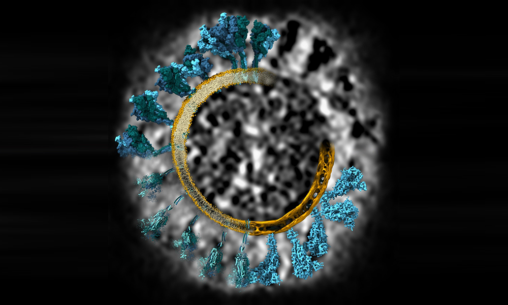
This stunning image shows SARS-CoV-2’s spike protein (blue) in gradually increasing detail, from initial experimental data to a final 3D model. Beata Turoňová, then a postdoc in the Beck group at EMBL Heidelberg, and Mateusz Sikora, a postdoc in the lab of Gerhard Hummer at the Max Planck Institute of Biophysics, prepared the image to illustrate their workflow. Additional details were revealed as the experiments proceeded, until the scientists arrived at a full 3D rendering of the spike protein – and subsequently a full 3D model of a single virus particle.
The black and white image at the top right is a slice from one of the cryo-electron tomograms – the experimental data on which all the following spike protein models are based. The scientists used these tomograms to calculate 3D maps of the spike protein and the viral membrane (yellow) to which it’s anchored. At first, only broad features were discernible (bottom right); then, the scientists were able to pinpoint the location of individual atoms (bottom left). Finally, the scientists used the data to render 3D models of all spike proteins on a single virus, some of which are shown at top left.
The image illustrates the results of a collaborative research project that found an unexpected flexibility of movement within the spike protein, and revealed an additional protective layer of sugar-like molecules on its surface. The project was carried out by a team of scientists at EMBL Heidelberg, the Max Planck Institute of Biophysics, the Paul-Ehrlich-Institut, and Goethe University Frankfurt; the results were published in Science in August 2020. To learn more about the process that led Beata and Mateusz to create this image, read their interview published on the Science Visuals blog.
Credit: Beata Turoňová/EMBL; Mateusz Sikora/MPI of Biophysics
If you have a stunning picture of your science, your lab or your site, you can submit it here.


