First, find your membrane
Collaboration between scientists reveals collaboration between lipids, in a study published in Cell Reports.

Last year, Rainer Pepperkok (Head of Core Facilities) showed that if you destroy the Golgi complex – the cell’s packaging centre – it gets rebuilt. “That’s fascinating!” says group leader Anne-Claude Gavin. “How does the cell first know that it is damaged, and that it has to build it again? And how does it know when to stop?” It’s just one of a multitude of examples Gavin can give you of instances where the cell needs a sensing mechanism associated to the membranes that partition off different compartments like the Golgi or the nucleus – not to mention the membrane that encompasses the cell itself.
Such sensing mechanisms involve, among others, proteins that sense and dock onto a specific membrane. But many of those proteins are known to bind to lipids which exist in many membranes. So how do they ‘know’ which particular membrane to bind to at a given time – for example, to start a bud that will develop into a new yeast cell?
Gavin’s lab developed a new approach to this question, together with the labs of Peer Bork and Jan Ellenberg. It stemmed from a brainstorming session with postdoc Antoine-Emmanuel Saliba, who under EMBL’s Interdisciplinary Postdoc Programme was affiliated to both the Gavin and Ellenberg groups. “We were dreaming up a way to measure these protein-lipid interactions using microfluidics, because Antoine-Emmanuel had experience in microfluidics. We were thinking about something that was visual,” she says. “So microfluidics, but with microscopy, using biochemical methods, and of course bioinformatics to process and analyse the data. Ivana Vonkova, a PhD student in my lab, developed the biochemical methods. Antoine-Emmanuel and Jan were the microfluidics and microscopy experts. Samy Deghou, from the Bork group, came up with innovative ideas so that we could integrate and visualise the huge amount of data you produce when you run 30 000 experiments.”
So microfluidics, but with microscopy, using biochemical methods, and of course bioinformatics to process and analyse the data.
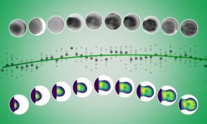
In their 30 000 experiments, the team investigated how so-called pleckstrin homology domains (PH domains) – the ‘hand’ that many proteins use to attach themselves to membranes – bind to different artificial membranes. PH domains are known to bind to a group of lipids called PIPs. But virtually every membrane in the cell contains PIPs, so how does a given protein bind only to one particular type of membrane, at a specific time? Gavin and colleagues created ‘bubbles’ of membrane made up of a PIP and another signalling lipid. By playing with the concentration of PIPs and combining them with different lipids, they created 10000 different scenarios in which they could measure how well PH domains bound. Each scenario was run three times, to ensure results weren’t due to chance.
They found that the degree and timing of binding are fine-tuned by additional lipids. PIP acts as a flag, saying ‘this is the membrane,’ and the presence of a second lipid pinpoints further: ‘this is the section of membrane to bind to now.’
For instance, when a yeast cell is about to reproduce, its cell membrane starts growing in one spot, creating a bud that eventually breaks loose as a separate cell. Some proteins with a PH domain are involved in this process by recognising a specific PIP called PI(4,5)P2 – which is present on all of the cell’s membrane. The EMBL scientists discovered that this PH domain binding is much stronger if another lipid, phosphatidylserine, is present. This explains how PH domains can drive yeast budding, since phosphatidylserine accumulates in the section of membrane that is set to bud out.
The EMBL scientists believe that other instances of protein-lipid binding on membranes likely also involve fine-tuning by a second lipid.
Could it be that you recognise one lipid in a thin membrane, another in a more flexible membrane?
Next, Gavin and colleagues would like to investigate how the physical properties of membranes influence this recognition process. “We know that the endoplasmic reticulum is thin and floppy, whereas the plasma membrane is thick and rigid, … Could it be that you recognise one lipid in a thin membrane, another in a more flexible membrane?” she wonders, adding: “We’d like to explore this, either using real membranes, which will be challenging, or by mimicking these properties in our artificial membranes.”


