EMBL Hamburg scientists have revealed how an unstructured protein traps cancer-promoting molecules
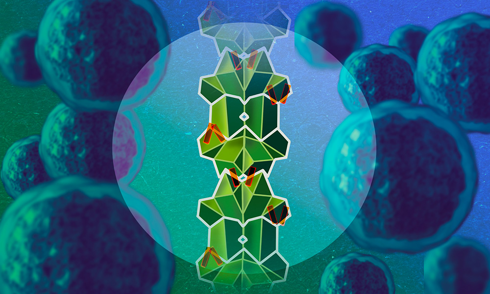
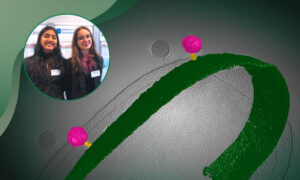
A visual metaphor depicting the unstructured protein RAI2 (parts of it visible in orange), as it disables the cancer-promoting CtBP protein (green) by stacking its molecules up into an elongated polymer which forms aggregates in cells. This process may play a role in how cancer progresses to become treatment resistant. Credit: Isabel Romero Calvo/EMBL
Summary
EMBL Hamburg scientists and collaborators discovered a new molecular mechanism through which an unstructured protein disables cancer-promoting molecules by gluing them together into a stack.
Unstructured proteins are essentially ‘invisible’ to structural biology techniques, which makes them challenging to study.
Data from human patient samples support the role of this mechanism in cancer progression. While the study mostly focused on prostate cancer, the mechanism likely plays a role in other types of cancer as well.
Each second of our lives, cells in our body grow and divide to ensure we stay healthy. However, this process has a dark side: if cell growth and divisions become excessive, that may cause cancer. To keep a safe balance, our cells are equipped with several molecular mechanisms to limit their own growth and division.
Their discovery, like a good novel, has a villain and a hero.
The ‘villain’ in this story is a group of proteins, called CtBPs (C-terminal binding proteins), which regulate the activity of several genes involved in cell growth and division. In various studies, CtBPs have been demonstrated to participate in promoting the development of cancer.
But the cell has its ways to harness the ‘villain’ – that’s where the ‘hero’ comes in: protein RAI2 (Retinoic Acid-Induced 2). RAI2 was only discovered recently, and even though it’s present in the cell in small amounts, it has been shown to play a role in preventing cancer metastasis. However, until now, it has been unclear how it achieves this.
The scientists have discovered that RAI2 has a remarkable ability to seize CtBP molecules and stack them together in a process called ‘polymerisation’. As they get piled up, the CtBP molecules form elongated aggregates in cancer cell lines, described as ‘nuclear foci’. Trapped and stacked by RAI2 in the aggregate, CtBP, the villain, becomes inactive.
“Polymerisation has become an emerging field of interest in the life sciences, but to our knowledge, it has not been described to date in the context of interfering with cancer progression,” said Matthias Wilmanns, Group Leader at EMBL Hamburg. “The discovery of this new mechanism of powerful target inhibition could open up new anti-cancer therapy directions, such as developing small molecules with potential or cancer target polymerisation.”
An invisible hero
To structural biologists like the Wilmanns Group, RAI2, the hero, is essentially an ‘invisible’ protein. While the majority of proteins, like origami, fold into 3D structures, RAI2 rather resembles a flexible band that keeps changing shape. Capturing such a wobbly protein in high resolution is impossible with the currently available structural biology techniques.
“You could compare it to trying to take a photo of a moving object at night – what you’ll get is a blurry image,” said Wilmanns.
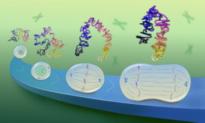
In this study, the only parts of RAI2 that could be visualised in the molecular model were the only parts that would stand still – the ‘sticky’ fragments through which RAI2 binds CtBPs. Each RAI2 molecule has two such fragments (depicted in orange in the images), which enable it, akin to adhesive tape, to glue two CtBP molecules together.
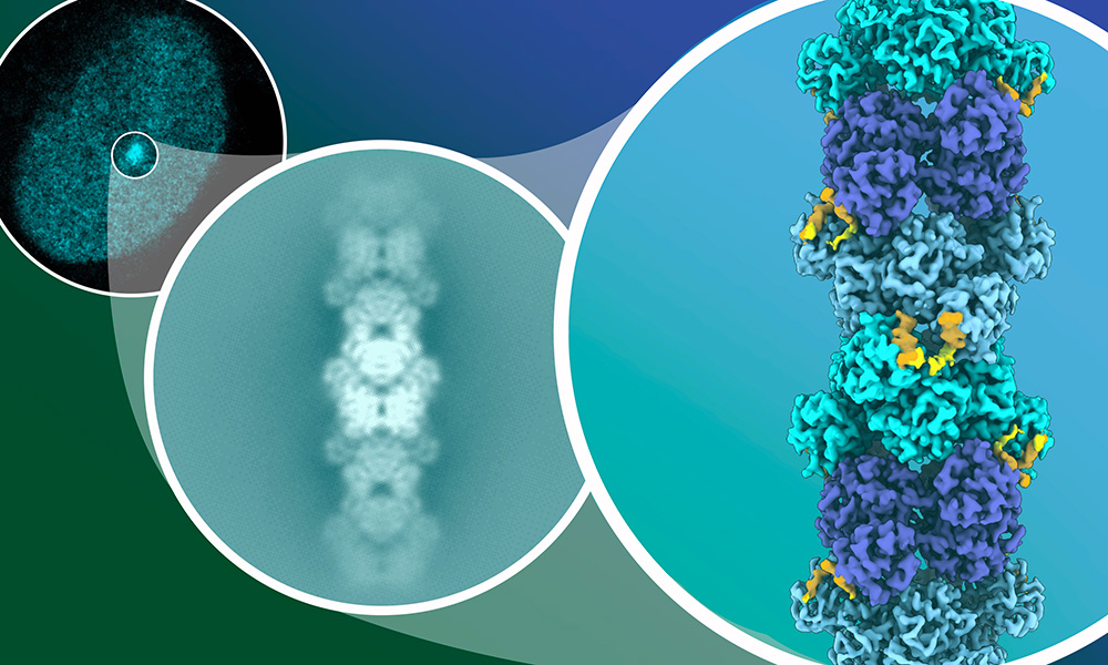
The scientists observed the CtBP-RAI2 polymers at different scales: at the cellular level, as aggregates, or ‘foci’, in the nucleus (upper left) and at the molecular level as a stack of molecules captured with cryo-electron microscopy (middle). The latter enabled them to create a detailed structural model (right). Credit: Isabel Romero Calvo/EMBL
“Wobbly proteins like RAI2 are challenging to work with, so this project took lots of hard work, focus, determination, and a ‘never give up’ attitude,” said Nishit Goradia, previously a postdoc in the Wilmanns Group, currently a fellow at the UKE. “We’ve revealed that RAI2 is a real dark horse: like other wobbly proteins, it’s quite understudied, but it holds true potential for inactivating cancer-promoting proteins. For me, this has been a career-defining project and I’m proud of this achievement.”
Wobbly protein in prostate cancer
To check if the new mechanism plays a role in human cancer, the scientists analysed, in addition to cancer cell lines, cancer samples from a diverse cohort of more than 100 patients of the UKE’s University Cancer Center Hamburg and Martini Clinic. They focused on prostate cancer, the second most common type of cancer in men and the third leading cause of cancer related death.
In the course of therapy, prostate cancer may develop resistance to treatment, resulting in very poor prognosis for the patients. However, it is not yet fully understood why some forms of prostate cancer develop into certain highly aggressive subtypes, and others don’t. RAI2 could play a key role here.
In both tumour cell lines and samples from patients, the scientists saw that the RAI2 levels were strongly reduced in more severe and treatment-resistant types of prostate cancer. In analyses in vitro, they saw that loss of RAI2 favours cellular processes that may lead to resistance to certain types of treatment.
“Even if we are not yet able to use this result therapeutically, it is a decisive step towards better understanding how a very aggressive subtype of prostate cancer develops,” said Gunhild von Amsberg, Professor of Uro-oncology at the University Cancer Center Hamburg and the Martini Clinic. “An important next step will be to transfer our findings to the clinic and thus identify patients who may be at risk at an early stage.”
Although the analysis focused on prostate cancer, the scientists suspect that the significance of their discovery maybe be applicable to other cancers too.
Molecular biology meets the clinics
The study is part of the long-standing collaboration between EMBL and UKE and showcases the benefits of combining both institutions’ complementary scientific approaches. While the UKE conducts a large spectrum of medical projects, in part based on patient data, EMBL contributes expertise in studying life across biological scales, including investigating disease mechanisms at the molecular and cellular levels.
“This work demonstrates the future perspective of collaboration between molecular biology and the clinics to unlock research ‘from molecules to patients’,” said Wilmanns.