27 October 2023
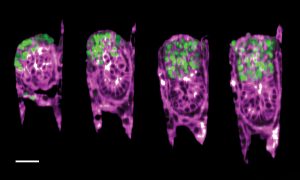
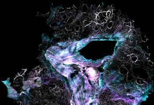
EMBL researchers have created an engineered uterus that allows a closer look at a mouse embryo’s development and its interactions with the uterine environment.
SCIENCE & TECHNOLOGY
2023
picture-of-the-weeksciencescience-technology
31 August 2023
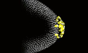
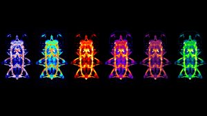
For a fruit fly embryo to develop correctly, key factors need to get to the right place at the right time – a journey that starts in the developing egg, as seen in this image from the Ephrussi Group at EMBL Heidelberg
SCIENCE & TECHNOLOGY
2023
picture-of-the-weeksciencescience-technology
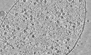
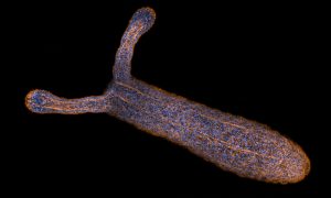
10 July 2023
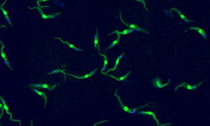
This single-celled organism the size of a dust particle is capable of causing deadly tropical diseases in both humans and livestock –Trypanosoma brucei, in an image by Luciano Dolce from EMBL.
LAB MATTERSSCIENCE & TECHNOLOGY
2023
lab-matterspicture-of-the-weekscience-technology
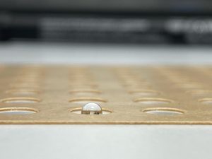
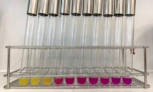
23 February 2023
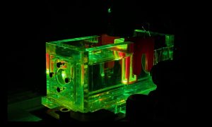
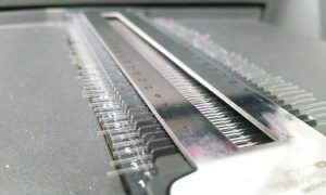
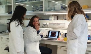
Researchers in the Prevedel Group use photoacoustic spectroscopy setup to test and optimise probes before their usage in mouse neuroscience.
LAB MATTERSSCIENCE & TECHNOLOGY
2023
lab-matterspicture-of-the-weekscience-technology
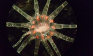
20 January 2023
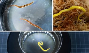
Tasmanian flatworms add to an EMBL researcher’s collection as she studies principles that control animal body size.
LAB MATTERSSCIENCE & TECHNOLOGY
2023
lab-matterspicture-of-the-weekscience-technology
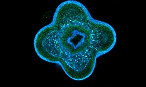
19 January 2023
Mucus present in the mouse colon can be visualised using Alcian blue staining, as imaged here by EMBL predoctoral fellow Linda Decker.
LAB MATTERSSCIENCE & TECHNOLOGY
2023
lab-matterspicture-of-the-weekscience-technology
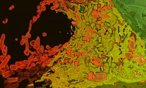
23 December 2022
Zebrafish embryos during gastrulation, a very early stage of development, to study the effect of temperature on vertebrate embryo development.
LAB MATTERSSCIENCE & TECHNOLOGY
2022
lab-matterspicture-of-the-weekscience-technology
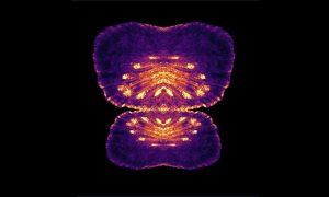
21 December 2022
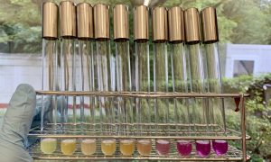
To identify which drugs disrupt bacterial envelope integrity, the Typas group uses a molecule called chlorophenyl red-β-D-galactopyranoside.
LAB MATTERSSCIENCE & TECHNOLOGY
2022
lab-matterspicture-of-the-weekscience-technology
31 August 2022
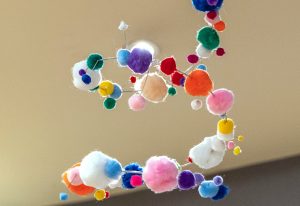
Physarum polycephalum, a single, giant cell containing tens of thousands of nuclei is large enough to be photographed with a phone.
SCIENCE & TECHNOLOGY
2022
picture-of-the-weekscience-technology
24 August 2022
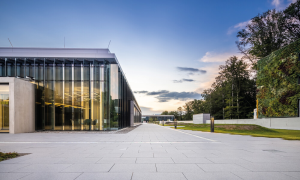
EMBL’s imaging centre makes advanced microscopy technologies accessible to the international scientific community.
LAB MATTERS
2022
lab-matterspicture-of-the-week
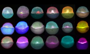
29 June 2021
As evocative as a constellation of planets, these egg cells within a mouse ovary are at different stages of maturity.
SCIENCE & TECHNOLOGY
2021
picture-of-the-weeksciencescience-technology
23 June 2021

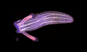
EMBL PhD student Anniek Stokkermans captured this side view of a Nematostella vectensis larva during this transition, using instrumentation in the Advanced Light Microscopy Facility at EMBL Heidelberg.
SCIENCE & TECHNOLOGY
2021
picture-of-the-weekscience-technology
15 June 2021
As perfect as a summer night sky, these nuclear pores help calibrate a customised super-resolution microscope in EMBL’s Ries group.
SCIENCE & TECHNOLOGY
2021
picture-of-the-weekscience-technology
8 June 2021
At EMBL, we have many dream teams – groups of individuals who support each other, innovate, and work together. One of those dream teams bridges two core facilities at EMBL Rome.
SCIENCE & TECHNOLOGY
2021
picture-of-the-weekscience-technology
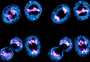
1 June 2021
Captured by EMBL postdoc Arina Rybina, these ‘nuclear twins’ are two daughter nuclei straight after division of a HeLa cell.
SCIENCE & TECHNOLOGY
2021
picture-of-the-weekscience-technology
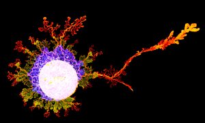
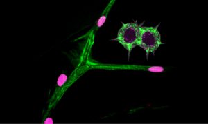
25 May 2021
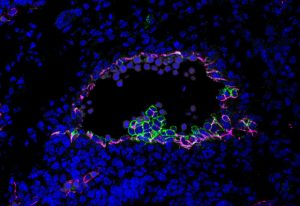
The Bernabeu Group aims to increase our knowledge of cerebral malaria, using in vitro engineered networks of human blood vessels and brain cells.
SCIENCE & TECHNOLOGY
2021
picture-of-the-weekscience-technology
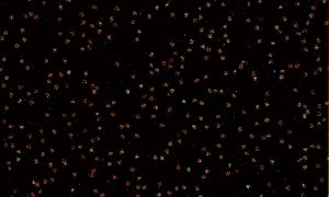
18 May 2021
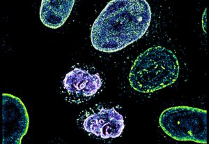
The EMBL Picture of the Week features a series of Jurkat T cells during different stages of the activation process.
SCIENCE & TECHNOLOGY
2021
picture-of-the-weekscience-technology
27 April 2021
A page from a biologist’s colouring book? EMBL’s new interior wall design? Not quite – a bunch of liver cells, grown in the lab so that scientists can learn about fatty liver disease, or steatosis.
SCIENCE & TECHNOLOGY
2021
picture-of-the-weekscience-technology
20 April 2021
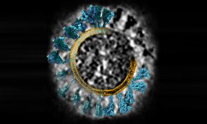
What does coronavirus’s spike protein look like in 3D? EMBL scientists and colleagues used cryo-electron tomography and molecular dynamics simulations to find out.
SCIENCE & TECHNOLOGY
2021
picture-of-the-weekscience-technology
13 April 2021
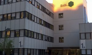
Throwback to June 2014: While EMBL Heidelberg’s main entrance is still in the dark, the well-known EMBL logo is already lit by the morning sun.
LAB MATTERS
2021
lab-matterspicture-of-the-week
6 April 2021
This image of a young Nematostella vectensis polyp shows two of the characteristic tentacles as well as the gaping mouth of the animal.
SCIENCE & TECHNOLOGY
2021
picture-of-the-weekscience-technology
30 March 2021
Scientists at EMBL Hamburg use droplets of protein solution to grow protein crystals. By exposing the crystals to X-rays, they are able to determine the protein’s molecular structure.
SCIENCE & TECHNOLOGY
2021
picture-of-the-weekscience-technology
23 March 2021

The cafeteria at EMBL Heidelberg now offers espresso and cappuccino with a more intense flavour and which are produced in a ‘green’ way.
LAB MATTERS
2021
lab-matterspicture-of-the-week
16 March 2021

EMBL’s site in Monterotondo has recently been blessed with blue skies and sunshine, leading to trees blooming early on campus.
LAB MATTERS
2021
lab-matterspicture-of-the-week
9 March 2021
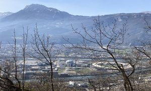
This photo shows the European Photon and Neutron (EPN) science campus where EMBL Grenoble is located. A delightful spring breeze in the air melts the last remaining patches of snow in the mountains.
LAB MATTERS
2021
lab-matterspicture-of-the-week
2 March 2021
Like caterpillars turning into beautiful butterflies, fruit fly larvae have to go through metamorphosis to finish their development. However, despite the fruit fly Drosophila melanogaster being one of the best studied model organisms in biology, comparatively little attention has been given to this…
SCIENCE & TECHNOLOGY
2021
picture-of-the-weekscience-technology
23 February 2021
Liang Xue used cryo-electron tomography to capture this detailed image of a Mycoplasma pneumoniae cell.
SCIENCE & TECHNOLOGY
2021
picture-of-the-weekscience-technology
16 February 2021
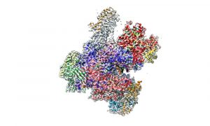
This week's Picture of the Week, which could also be a masterpiece of modern art, shows the enzyme RNA polymerase III, an assembly of 17 individual proteins combined into this complex structure.
SCIENCE & TECHNOLOGY
2021
picture-of-the-weekscience-technology
9 February 2021
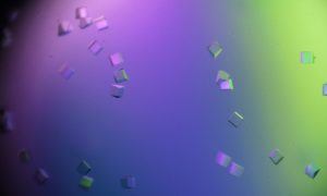
The regular structures of crystals are a source of inspiration and fascination to us humans. While the crystals in this picture were not grown in nature, but instead by Petra Drncova from EMBL Grenoble, they share the same attributes as those found in nature.
SCIENCE & TECHNOLOGY
2021
picture-of-the-weekscience-technology
2 February 2021
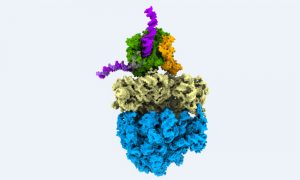
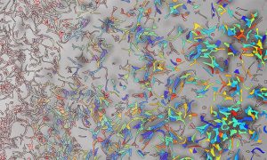
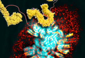
This colourful image shows biological information flow in action: It’s a supramolecular assembly of DNA, RNA and proteins, observed directly inside a bacterial cell while turning genetic information into protein.
SCIENCE & TECHNOLOGY
2021
picture-of-the-weekscience-technology
26 January 2021
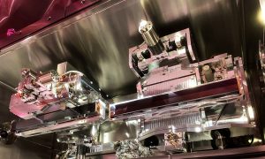
Scientists at EMBL Hamburg use specially designed mirrors to reflect and focus X-ray beams onto tiny crystals made of proteins or other biological molecules.
SCIENCE & TECHNOLOGY
2021
picture-of-the-weekscience-technology
19 January 2021
It’s almost a year since the coronavirus outbreak was declared a pandemic, affecting all our lives. While the virus continues its grip on the world, scientists are understanding it better and better, increasing our knowledge about it and opening up new ways to fight it.
SCIENCE & TECHNOLOGY
2021
picture-of-the-weekscience-technology
12 January 2021
Structural biologists want to study proteins at the atomic level. The device shown in this Picture of the Week is essential for this.
SCIENCE & TECHNOLOGY
2021
picture-of-the-weekscience-technology
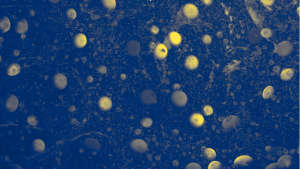
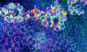
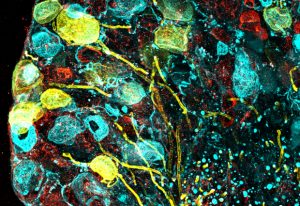
5 January 2021
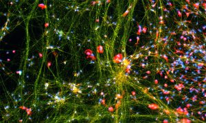
Fluorescent dyes light up a cellular community of neurons and brain immune cells (microglia), which were derived from stem cells.
SCIENCE & TECHNOLOGY
2021
picture-of-the-weekscience-technology
22 December 2020
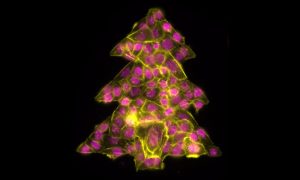
It is that time of year to get into the holiday spirit, prepare for some time at home and relax after a strange and stressful year. Even the cells in our Picture of the Week are getting into the holiday spirit, forming this colourful Christmas tree.
SCIENCE & TECHNOLOGY
2020
picture-of-the-weekscience-technology
15 December 2020
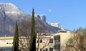
The first snowflakes have fallen on the Vercors Mountains, right behind the EMBL Grenoble building, and an almost full moon is showing up during a sunny morning.
LAB MATTERS
2020
lab-matterspicture-of-the-week
8 December 2020
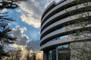
With the external scaffolding removed, another step in the construction of the EMBL Imaging Centre is complete. Now we get a first glimpse of the final look of this stunning building.
LAB MATTERS
2020
lab-matterspicture-of-the-week
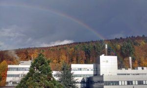
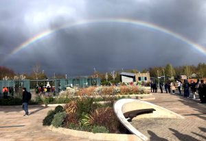
1 December 2020
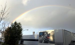
Hamburg hosts one of EMBL’s six sites. The city is notorious for its windy and rainy weather. Lots of rain also means a high chance of beautiful rainbows!
LAB MATTERS
2020
lab-matterspicture-of-the-week
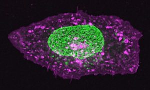
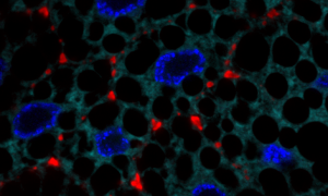
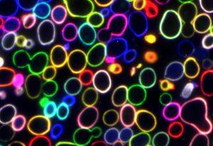
24 November 2020
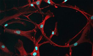
Studying cancers means also knowing what healthy cells look like. In this case, mesenchymal stromal cells (MSCs) from healthy bone marrow are a bit ‘loopy’.
SCIENCE & TECHNOLOGY
2020
picture-of-the-weekscience-technology
17 November 2020
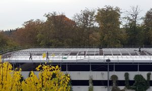
Despite the cold autumn weather, workers are busy on the rooftop of the parking garage at EMBL Heidelberg. The 2176 m² rooftop is getting transformed into a combination of a green roof and a photovoltaic plant. The planted green roof will retain rainwater, while the solar panels – installed in…
LAB MATTERS
2020
lab-matterspicture-of-the-week
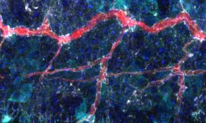
10 November 2020
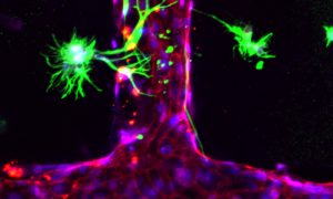
To help understand cerebral malaria the Bernabeu group has created in vitro engineered networks of human blood vessels.
SCIENCE & TECHNOLOGY
2020
picture-of-the-weekscience-technology
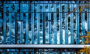
3 November 2020
On autumn days without Heidelberg’s characteristic fog, the woods present themself in beautiful colours. You may even capture a rainbow.
SCIENCE & TECHNOLOGY
2020
picture-of-the-weekscience-technology
27 October 2020
The nucleus of this cell fluoresces in bright green thanks to GFP-labelled nucleoporin proteins. EMBL scientists use engineered nucleoporins as 3D reference standards to improve super-resolution microscopy.
SCIENCE & TECHNOLOGY
2020
picture-of-the-weekscience-technology
20 October 2020
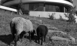
In 2012, EMBL used ecological lawn mowers to keep the grass short around the Advanced Training Centre at EMBL Heidelberg.
LAB MATTERS
2020
lab-matterspicture-of-the-week
13 October 2020
Sea anemones are amazing creatures. Despite their plant-like appearance and their tendency to remain fixed in one spot, they are actually animals.
SCIENCE & TECHNOLOGY
2020
picture-of-the-weekscience-technology
6 October 2020
We’ve all had wounds at certain times in our lives. But they heal due to the self-repairing mechanisms in the body.
SCIENCE & TECHNOLOGY
2020
picture-of-the-weekscience-technology
29 September 2020
To study the effect of commonly used drugs on bacterial envelopes, EMBL scientists applied a biochemical assay using a colour reaction. The deeper the red, the stronger the disruptive effect of the drug.
SCIENCE & TECHNOLOGY
2020
picture-of-the-weekscience-technology
22 September 2020
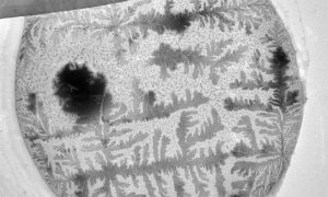
How does your crystal garden grow? EMBL's Electron Microscopy Core Facility was able to capture this garden of blooming crystals as they studied mosquito reproductive cells.
SCIENCE & TECHNOLOGY
2020
picture-of-the-weekscience-technology
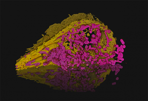
15 September 2020
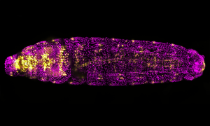
Not just another pretty fruit fly. This magenta and golden drosophila larva is lit up with a fluorescent molecule to help researchers study heart formation.
SCIENCE & TECHNOLOGY
2020
picture-of-the-weekscience-technology
8 September 2020
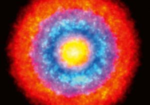
While this may seem like a nebula made up of interstellar clouds of dust and ionised gases, this image isn’t of a galaxy beyond the Milky Way.
SCIENCE & TECHNOLOGY
2020
picture-of-the-weekscience-technology
1 September 2020
Those heart-shaped cells aren't just for show. They help tell the story of two proteins working together
SCIENCE & TECHNOLOGY
2020
picture-of-the-weekscience-technology
25 August 2020
Beautiful flashes of blue colour help light the way for researchers to study cells in fruit fly larva that provide oxygen to tissues.
SCIENCE & TECHNOLOGY
2020
picture-of-the-weekscience-technology
18 August 2020
Despite their ghostly appearance, these are very real cell nuclei infected with Influenza A virus – the only influenza virus known to cause pandemics.
SCIENCE & TECHNOLOGY
2020
picture-of-the-weekscience-technology
11 August 2020
This image is a composite of lateral pentascolopidial organs, a wing imaginal disc pouch, and an epithelial wound in a Drosophila larva. The organs are arranged here like eyelashes. Cells surrounding an epidermal wound appear as the iris and pupil of this artistic eye.
SCIENCE & TECHNOLOGY
2020
picture-of-the-weekscience-technology
4 August 2020
This group of cells represents an interesting example of organ formation where cells simultaneously move and change their shapes in a highly coordinated manner.
SCIENCE & TECHNOLOGY
2020
picture-of-the-weekscience-technology
28 July 2020
Bacterial cells are embedded in microfluidic droplets in oil. The fluorescence indicates the presence of the targeted DNA strain with the help of a characteristic DNA sequence.
SCIENCE & TECHNOLOGY
2020
picture-of-the-weekscience-technology
21 July 2020
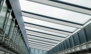
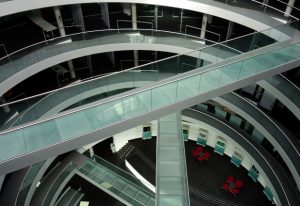
The image shows one of the four rows of roof lights above the atrium, which is the main public space of the Imaging Centre.
SCIENCE & TECHNOLOGY
2020
picture-of-the-weekscience-technology
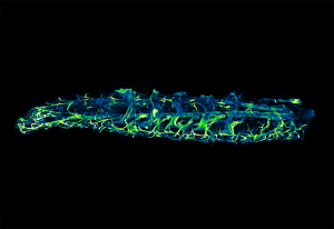
14 July 2020
The image shows a larva of Platynereis dumerilii, a marine worm. The image here was produced by Constantin Pape, a visiting predoctoral fellow in the Kreshuk group at EMBL Heidelberg.
SCIENCE & TECHNOLOGY
2020
picture-of-the-weekscience-technology
7 July 2020
EMBL brings together more than 1700 people from all over the world, from a variety of academic and cultural backgrounds. This creates an environment in which there is constant exchange of both scientific knowledge and cultural heritage. While it seems obvious that EMBL, as an international…
LAB MATTERS
2020
lab-matterspicture-of-the-week
30 June 2020
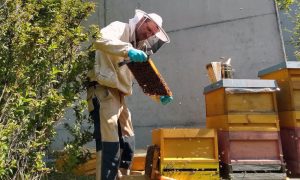
Local beekeeper Jörg Staffel has set up three bee colonies on the grass slope in front of the ATC building.
LAB MATTERS
2020
lab-matterspicture-of-the-week
23 June 2020
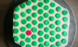
Elisa Kreibich from EMBL’s Krebs group baked an EMBL cake for the farewell of Alexandra Martitz, the first master student in their lab.
LAB MATTERS
2020
lab-matterspicture-of-the-week
16 June 2020
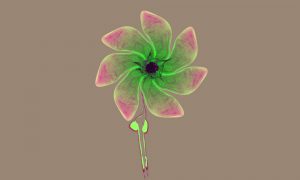
In this composite image, visual artist Mona Kakanj assembled three different biological structures in fly larvae into a flower. The original images were taken as part of a research project by Parisa Kakanj in Maria Leptin’s group.
SCIENCE & TECHNOLOGY
2020
picture-of-the-weekscience-technology
9 June 2020
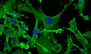
This image shows mouse embryonic fibroblasts (MEFs), their cell skeletons (green) and nuclei (blue) under a confocal microscope, photographed by Julia Hansen in the lab of Matthieu Boulard at EMBL Rome.
SCIENCE & TECHNOLOGY
2020
picture-of-the-weekscience-technology
2 June 2020
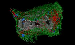
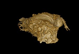
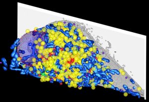
This image is a 3D-rendering of the automatic segmentation of a HeLa cell, made by Julian Hennies from the Schwab team at EMBL Heidelberg.
SCIENCE & TECHNOLOGY
2020
picture-of-the-weekscience-technology
26 May 2020
In the Leptin Group, Eva Hasel investigates the innate immune system in Japanese rice fish.
SCIENCE & TECHNOLOGY
2020
picture-of-the-weekscience-technology
19 May 2020
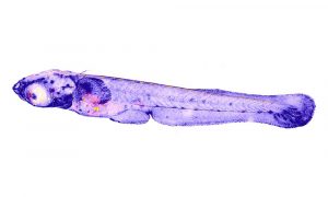
The image shown here, taken by Anniek Stokkermans from EMBL’s Ikmi Group, shows a larva of Nematostella vectensis.
SCIENCE & TECHNOLOGY
2020
picture-of-the-weekscience-technology
12 May 2020
This image of the starlet sea anemone was taken by Petrus Steenbergen using a stereoscope.
SCIENCE & TECHNOLOGY
2020
picture-of-the-weekscience-technology
6 May 2020
The image shown here, taken by Daniel Rios-Barrera from the Leptin Group shows the cells of the early wing tissue of the fly during larval development.
SCIENCE & TECHNOLOGY
2020
picture-of-the-weekscience-technology
28 April 2020
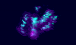
In human cells, the genetic material is packaged into 23 different DNA molecules, the chromosomes. Each chromosome is present in two copies, one inherited from the paternal sperm, and the other from the maternal egg. During most of the cell’s life, chromosomes take the shape of long,…
SCIENCE & TECHNOLOGY
2020
picture-of-the-weekscience-technology
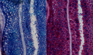
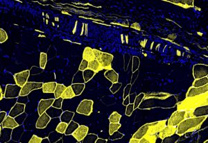
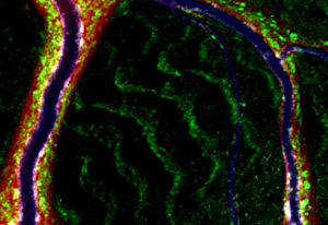
21 April 2020
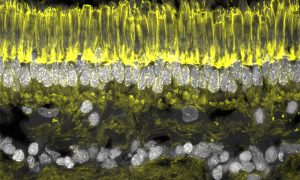
This Picture of the Week shows a stained cryosection of a retina – the light-sensitive part of the eye – of an ancient fish, the lamprey.
SCIENCE & TECHNOLOGY
2020
picture-of-the-weekscience-technology
14 April 2020
What may look like a branch of a tree with the first flower buds emerging after winter are, in fact, tracheal cells of a fruit fly larva and their nuclei.
SCIENCE & TECHNOLOGY
2020
picture-of-the-weekscience-technology
7 April 2020
EMBL is all about exciting science, through which we aim to achieve a fundamental understanding of biological processes.
SCIENCE & TECHNOLOGY
2020
picture-of-the-weekscience-technology
31 March 2020
Paola Bertucci, from the Arendt Group at EMBL Heidelberg, studies the evolution of Platynereis dumerilii – a species of annelid polychaete worm.
SCIENCE & TECHNOLOGY
2020
picture-of-the-weekscience-technology
24 March 2020
The iconic ATC – celebrating its 10 year anniversary this year – reflects the blue sky, the clouds and the rays of the Sun.
LAB MATTERS
2020
lab-matterspicture-of-the-week
17 March 2020
The medaka shown in this Picture of the Week was captured by Eva Hasel, a postdoc in the Leptin group at EMBL Heidelberg.
SCIENCE & TECHNOLOGY
2020
picture-of-the-weekscience-technology
10 March 2020

This image, taken by Raphaël Cohen-Aberdam from the Cipriani team, shows that the weather for the Grenoble Lab Ski Day 2020 was just perfect!
LAB MATTERS
2020
lab-matterspicture-of-the-week
3 March 2020
Colder winter weather has finally arrived EMBL-EBI. It’s the first time the site has seen snow this winter.
LAB MATTERS
2020
lab-matterspicture-of-the-week
25 February 2020
In the Trivedi Group at EMBL Barcelona, Krisztina Arató and Jia Le Lim study the early development of zebrafish embryos.
SCIENCE & TECHNOLOGY
2020
picture-of-the-weekscience-technology
18 February 2020
In this image, Julian Hennies from the Schwab Team has reconstructed the 3D structure of a human cell's organelles.
SCIENCE & TECHNOLOGY
2020
picture-of-the-weekscience-technology
11 February 2020
This image shows the tracheal system of a live fruit fly larva. Daniel Rios from the Leptin Group and Dimitri Kromm from the Hufnagel Group used this advanced microscope to investigate the dynamics of tracheal cells during development.
SCIENCE & TECHNOLOGY
2020
picture-of-the-weekscience-technology
4 February 2020
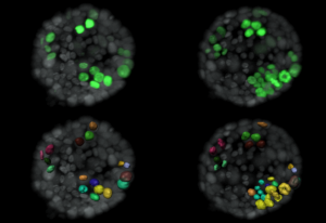
Morgan Oatley and her colleagues in Christophe Lancrin’s group investigated how haematopoietic stem cells emerge from the endothelium in developing mouse embryos.
SCIENCE & TECHNOLOGY
2020
picture-of-the-weekscience-technology
28 January 2020
Muzamil Majid Khan, a postdoc in the Pepperkok team at EMBL Heidelberg, studied the piece of tissue visible in this image.
SCIENCE & TECHNOLOGY
2020
picture-of-the-weekscience-technology
21 January 2020
The image shown here is a 3D-rendering created by Julian Hennies from the Schwab team.
SCIENCE & TECHNOLOGY
2020
picture-of-the-weekscience-technology
14 January 2020
This image has been composed from thousands of individual super-resolution microscopy images. It was created by Markus Mund in the Ries Group.
SCIENCE & TECHNOLOGY
2020
picture-of-the-weekscience-technology
7 January 2020
What might also be an artistic representation of a scattered bowl of Skittles is actually a 3D reconstruction of a high-pressure frozen HeLa cell.
SCIENCE & TECHNOLOGY
2020
picture-of-the-weekscience-technology
31 December 2019
Breast cancer is the second leading cause of cancer-related deaths in women. It is so deadly because tumours often return after successful cancer treatment. This recurrence is caused by individual dormant cancer cells remaining inside the breast. These cells can develop into active cancer cells…
SCIENCE & TECHNOLOGY
2019
picture-of-the-weekscience-technology
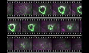
24 December 2019
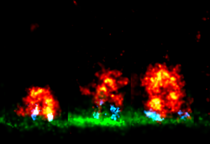
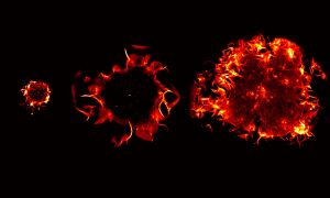
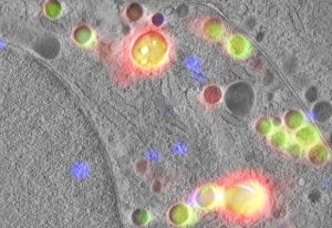
What looks like a photo-series of an explosive eruption are actually uptaking proteins, captured by Markus Mund from the Ries Group at EMBL Heidelberg. The images were made in an attempt to learn how the different proteins that take up molecules into the cells via endocytosis – the cellular…
SCIENCE & TECHNOLOGY
2019
picture-of-the-weekscience-technology
17 December 2019
DNA is present in each cell of our body. If all the DNA from one human cell was removed and aligned in a single strand, it would in theory add up to a total length of about two metres. In order to fit into the nucleus of a cell, DNA has to be compressed by […]
SCIENCE & TECHNOLOGY
2019
picture-of-the-weekscience-technology
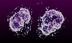
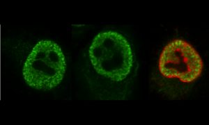
10 December 2019
This picture of the week, taken by Arina Rybina in the Ellenberg group at EMBL Heidelberg, shows a high-resolution 3D microscopy image of living human cells: HeLa cells. In this fascinating fluorescing microspace, two newly formed daughter nuclei are captured to study the assembly of nuclear pore…
SCIENCE & TECHNOLOGY
2019
picture-of-the-weekscience-technology
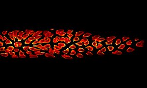
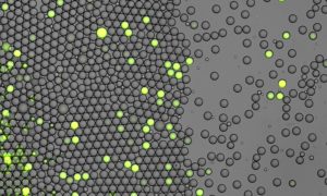
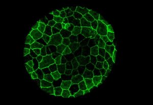
3 December 2019
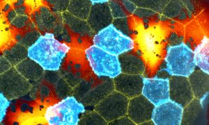
This beautiful mosaic of mostly hexagonal cells is the outer skin layer of a zebrafish larva as seen under a microscope. Each skin cell exhibits a unique pattern of actin ridges. Actin is a family of globular multifunctional proteins found in almost all eukaryotic cells. Actin forms microfilaments,…
SCIENCE & TECHNOLOGY
2019
picture-of-the-weekscience-technology
26 November 2019
The hormone insulin helps to remove sugar from the blood after a meal. This is important, as in the long term high blood sugar levels damage our bodies. Diabetes of type 1 or type 2 is a direct consequence of a failure to produce sufficient insulin or to release it from the cells in which […]
SCIENCE & TECHNOLOGY
2019
picture-of-the-weekscience-technology
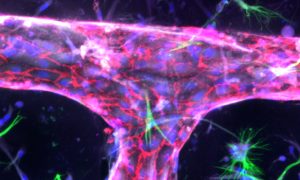
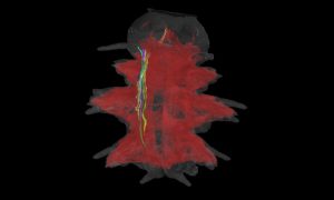
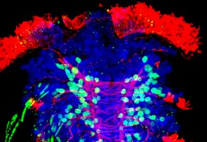
19 November 2019
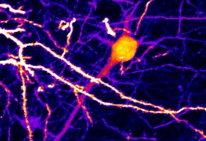
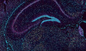
The brain is the most complex organ in the human body. Yet despite it being the organ that makes us conscious beings – and despite the fact that researchers have been studying it for generations – it’s still a constant source of surprise. To help lift the veil on some of its mystery, Lina…
SCIENCE & TECHNOLOGY
2019
picture-of-the-weekscience-technology
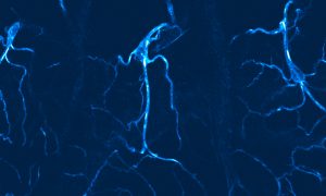
5 November 2019
Traditionally, we talk about having five senses: sight, hearing, taste, smell and touch. In reality, our bodies are capable of much more. Sitting right under our skin are a variety of sensory neurons, which are specialised in detecting light touch, pain, temperature, itch or the body’s position.…
SCIENCE & TECHNOLOGY
2019
picture-of-the-weekscience-technology
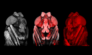
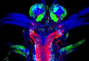
29 October 2019
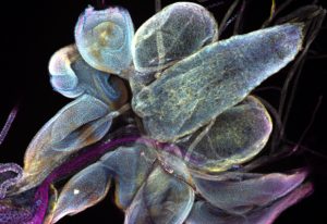
The three bluish blobs shown in the top right corner of this image may not resemble the sphere of noodles that is the human brain, but they are still essential – at least for the fruit fly. This Picture of the Week shows the brain lobes of Drosophila. It’s an insect so tiny and so […]
SCIENCE & TECHNOLOGY
2019
picture-of-the-weekscience-technology
22 October 2019
Is it a fungus or a strange plant? Actually it’s the larval form of Platynereis – a group of marine ringed worms. Scientists have been using them in their studies for the past 70 years, and they are among the preferred lab organisms. They are easy to keep in the lab, and under temperature and…
SCIENCE & TECHNOLOGY
2019
picture-of-the-weekscience-technology
15 October 2019
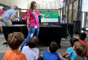
EMBL not only produces excellent science and innovative technologies; it also shares its knowledge and experience with partners from around the world – and with the public. As well as offering teacher training in the European Learning Laboratory for the Life Sciences, science movie nights, and…
LAB MATTERS
2019
lab-matterspicture-of-the-week
8 October 2019
Despite missing the characteristic stripes one would expect from a zebra – or a zebrafish – the fractals in this Picture of the Week show a zebrafish; or at least some cells in a zebrafish embryo, a few hours after fertilisation. Zebrafish are not only popular aquarium fish, they are also an…
SCIENCE & TECHNOLOGY
2019
picture-of-the-weekscience-technology
1 October 2019
Model organisms are species that are studied extensively to understand particular biological phenomena and processes, with the expectation that discoveries made in the model organism will provide insight into the workings of other organisms. The small marine ringed worm Platynereis dumerilii gained…
SCIENCE & TECHNOLOGY
2019
picture-of-the-weekscience-technology
25 September 2019
Fruit flies have something that we don’t have: they produce a protein called dumpy. This protein is the largest created by insects, and is comparable in size to the largest human protein – titin. While titin is vital for our muscle function, dumpy connects the soft cells of the insect’s…
SCIENCE & TECHNOLOGY
2019
picture-of-the-weekscience-technology
17 September 2019
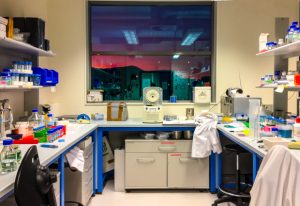
It’s evening and the Sun is setting over the mountains surrounding the city of Grenoble – home to one of EMBL’s six sites – bathing the mountaintops in fiery red light. The Picture of the Week, taken by Zuzanna Kaczmarska shows the lab she worked in after a long and busy day. Bottles…
LAB MATTERS
2019
lab-matterspicture-of-the-week
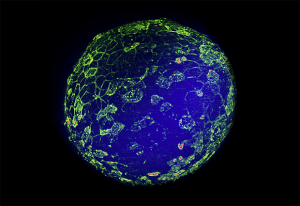
10 September 2019
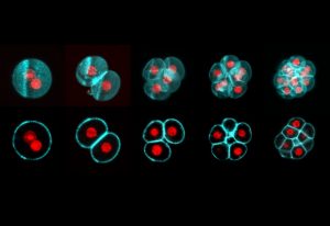
All mammalian life starts with the fusion of egg and sperm, resulting in the creation of a single cell called a zygote. This develops into an embryo through a series of cell divisions, in which the number of cells doubles at each step. Todays’ Picture of the Week was taken by Manuel Eguren of the…
SCIENCE & TECHNOLOGY
2019
picture-of-the-weekscience-technology
3 September 2019
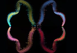
This gorgeous image of a stained adult marine worm was created by former EMBL postdoc Hernando Martinez using structured microscopy. The worm itself was captured during plankton extraction off the coast of Sweden. There are over 10 000 species of these swimming worms, and they have adapted to every…
SCIENCE & TECHNOLOGY
2019
picture-of-the-weekscience-technology
27 August 2019
The most basic building blocks of life are the biological molecules in our cells. While these molecules are too small to see with most microscopes, they have incredibly complex and beautiful structures. Therefore, the Protein Data Bank in Europe (PDBe), The Art Society CANTAB and The Art Society…
LAB MATTERS
2019
lab-matterspicture-of-the-week
20 August 2019
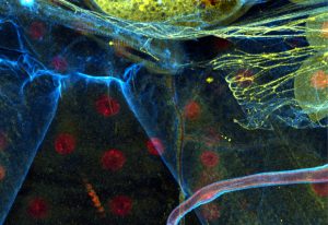
Today’s picture of the week is not only a colourful one, it is also a snapshot of the vast number of shapes that the cells inside an animal body can adopt. How this variety comes about is investigated in the Leptin group at EMBL Heidelberg. To understand the shapes of the cells in fruit fly…
SCIENCE & TECHNOLOGY
2019
picture-of-the-weekscience-technology
13 August 2019
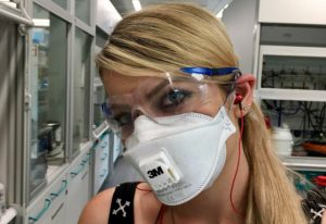
Working in a lab very often requires some kind of protection: gloves, safety goggles, lab coat, hearing protection. Sandra – now at BASF in Ludwigshafen – had fun getting ready for her work in the medicinal chemistry lab at EMBL! Here, she is about to grind potassium permanganate and copper…
PEOPLE & PERSPECTIVES
2019
people-perspectivespicture-of-the-week
6 August 2019
Low blood pressure (hypotension) or high blood pressure (hypertension) are risk factors for many diseases and affect more than 20% of the global population. How blood pressure is regulated is part of the research done in the Heppenstall group at EMBL Rome. In today’s Picture of the…
SCIENCE & TECHNOLOGY
2019
picture-of-the-weekscience-technology
30 July 2019
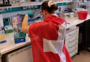
The 1700 people working at EMBL’s six sites come from more than 80 different countries. Many of them haven’t come straight from their home country to one of the EMBL sites but have also lived in other countries in between. While all of them are proud to work at EMBL they are also proud of…
PEOPLE & PERSPECTIVES
2019
people-perspectivespicture-of-the-week
23 July 2019
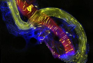
Every single moment of our life we use our muscles – most of the time without even thinking about it. Some muscles, like our heart, we cannot even control at all. How our brain communicates with our muscles is still not fully understood. The communication between our brain and our skeletal…
SCIENCE & TECHNOLOGY
2019
picture-of-the-weekscience-technology
16 July 2019
This colourful picture, taken by EMBL postdoc Arina Rybina using a confocal fluorescence microscope, shows human cells in the process of cell division. Eventually, each mother cell brings into existence two identical daughter cells. To visualise the process by light microscopy, different cell…
SCIENCE & TECHNOLOGY
2019
picture-of-the-weekscience-technology
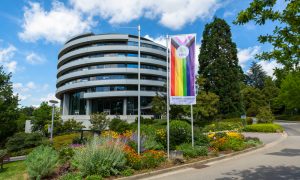
9 July 2019
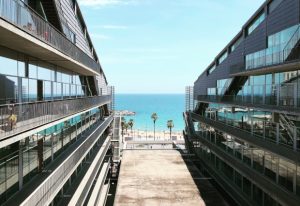
EMBL has six sites in Europe and the newest is EMBL Barcelona. It opened in October 2017 and is still growing. Eventually, it will be home to eight research groups, all of them working to discover how tissues and organs function and develop. EMBL Barcelona is located in the Barcelona Biomedical…
LAB MATTERS
2019
lab-matterspicture-of-the-week
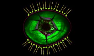
2 July 2019
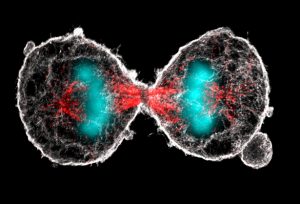
What looks like a pair of scary alien eyes is actually the final stage in the duplication of a cell. Cell duplication is preceded by a process called mitosis, in which the replicated chromosomes are separated into two new nuclei. Mitosis is the prerequisite for a cell to divide into two identical…
SCIENCE & TECHNOLOGY
2019
picture-of-the-weekscience-technology
25 June 2019
EMBL is an intergovernmental organisation, currently supported by 26 member states, one prospect and two associate member states. There are more than 1700 people working at EMBL, who come from more than 80 countries, creating a multicultural environment. EMBL also operates from six sites in Europe:…
LAB MATTERS
2019
lab-matterspicture-of-the-week
18 June 2019
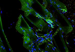
Have you ever wondered what reflex testing is about? Why does your doctor tap the space below your knee with a hammer to see if your leg kicks forward? At the centre of this involuntary reaction is the muscle spindle, of which you can see a close-up in today’s Picture of the Week. Muscle spindles…
LAB MATTERS
2019
lab-matterspicture-of-the-week
11 June 2019
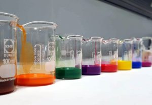
Laboratories all over the world are often chaotic, a bit messy and look grey and unwelcoming. Not so in this lab, pictured by EMBL staff member Emily Savage. The differently coloured fluids, arranged in a row, bring vivid colours into the more subdued environment of the lab. The picture was taken…
LAB MATTERS
2019
lab-matterspicture-of-the-week
4 June 2019
Most of us love brewer’s yeast, or at least the food that it’s helped us to produce since ancient times. Without Saccharomyces cerevisiae (its Latin name) we couldn’t enjoy wine, beer or most types of bread. Besides its role in food production, S. cerevisiae is also an important model…
LAB MATTERS
2019
lab-matterspicture-of-the-week
28 May 2019

EMBL’s sites provide spectacular views, such as this fiery sunset at the Wellcome Genome Campus in Hinxton. The campus, in the heart of the Cambridgeshire countryside, is home to several institutes and organisations working on genomics and computational biology. Among them is EMBL’s European…
LAB MATTERS
2019
lab-matterspicture-of-the-week
21 May 2019
This image – resembling a network of rivers and canals – actually shows the tracheal tip cell of a fruit fly. Fruit flies are heavily used in research and they are a common model organism in developmental biology. Researchers at EMBL use the larvae of fruit flies to study tracheal cell…
SCIENCE & TECHNOLOGY
2019
picture-of-the-weekscience-technology
14 May 2019
EMBL is a world-leading organisation for life science research. Its scientists work in diverse research fields spanning the whole of molecular biology. While the molecules the researchers are working on are often microscopic and impossible to see with the naked eye, one research topic clearly…
SCIENCE & TECHNOLOGY
2019
picture-of-the-weekscience-technology
7 May 2019
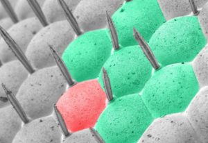
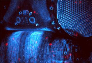
The hexagons visible in this Picture of the Week are the eyes of an ordinary housefly, visualised with a scanning electron microscope. Former staff member Anna Steyer, who captured this brilliant image, has coloured seven of the receptor areas of the eye to create a stylised version…
LAB MATTERS
2019
lab-matterspicture-of-the-week
24 May 2012
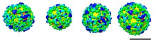
These spheres may look almost identical, but subtle differences between them revealed a molecular version of the robots from Transformers. Each sphere is a vesicle, a pod that cells use to transport materials between different compartments. The images, produced by Marco Faini from John…
SCIENCE & TECHNOLOGY
2012
picture-of-the-weekscience-technology
20 March 2012
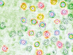
As spring arrives, flowers seem to bloom everywhere – even under the microscopes at the European Molecular Biology Laboratory (EMBL) in Heidelberg, Germany. But the ‘flowers’ in this picture actually help an animal, not a plant, to pass on its genes. The image, which has been false-coloured…
SCIENCE & TECHNOLOGY
2012
picture-of-the-weekscience-technology
No results found