5 October 2023
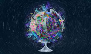
A new method developed by EMBL scientists can help us identify and investigate plankton species in field samples with greater speed, accuracy, and resolution than ever possible before.
SCIENCE & TECHNOLOGY
2023
sciencescience-technology
5 October 2021
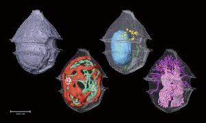
EMBL scientists and colleagues have developed an interactive atlas of the entire marine worm Platynereis dumerilii in its larval stage. The PlatyBrowser resource combines high-resolution gene expression data with volume electron microscopy images.
SCIENCE & TECHNOLOGY
2021
sciencescience-technology
22 September 2020
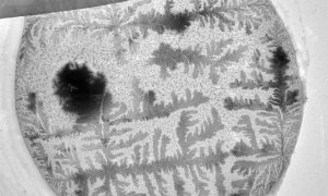
How does your crystal garden grow? EMBL's Electron Microscopy Core Facility was able to capture this garden of blooming crystals as they studied mosquito reproductive cells.
SCIENCE & TECHNOLOGY
2020
picture-of-the-weekscience-technology
No results found