4 January 2023
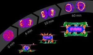
EMBL Heidelberg researchers and their collaborators reveal how the nuclear pore complex, one of the biggest molecular machines in eukaryotic cells, is assembled one protein at a time.
SCIENCE & TECHNOLOGY
2023
sciencescience-technology
5 July 2022
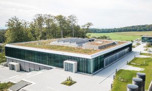
Creating a cutting-edge facility for the global life science community doesn't happen overnight. We spoke to some of those who worked to turn this dream into a reality.
CONNECTIONS
12 November 2021
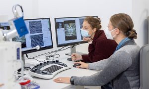
Correlative microscopy service enables PhD student from Switzerland to study structure and location of proteins cells use to communicate.
LAB MATTERSSCIENCE & TECHNOLOGY
2021
lab-mattersscience-technology
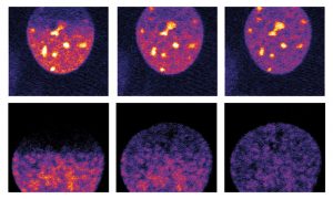
27 October 2020
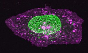
The nucleus of this cell fluoresces in bright green thanks to GFP-labelled nucleoporin proteins. EMBL scientists use engineered nucleoporins as 3D reference standards to improve super-resolution microscopy.
SCIENCE & TECHNOLOGY
2020
picture-of-the-weekscience-technology
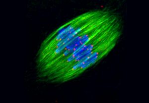
18 August 2011
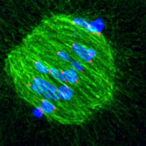
When an egg cell is being formed, the cellular machinery which separates chromosomes is extremely imprecise at fishing them out of the cell’s interior, scientists at the European Molecular Biology Laboratory (EMBL) in Heidelberg, Germany, have discovered. The unexpected degree of trial-and-error…
SCIENCE & TECHNOLOGY
2011
sciencescience-technology
23 January 2011
The sight of a researcher sitting at a microscope for hours, painstakingly searching for the right cells, may soon be a thing of the past, thanks to new software created by scientists at the European Molecular Biology Laboratory (EMBL) in Heidelberg, Germany. Presented today in Nature Methods, the…
SCIENCE & TECHNOLOGY
2011
sciencescience-technology
2 December 2010
From microscopy to computer tomography (CT) scans, imaging plays an important role in biological and biomedical research, but obtaining high-quality images often requires advanced technology and expertise, and can be costly. Euro-BioImaging, a project which launches its preparatory phase today,…
SCIENCE & TECHNOLOGY
2010
sciencescience-technology
1 April 2010
Name a human gene, and you’ll find a movie online showing you what happens to cells when it is switched off. This is the resource that researchers at the European Molecular Biology Laboratory (EMBL) in Heidelberg, Germany, and their collaborators in the Mitocheck consortium are making freely…
SCIENCE & TECHNOLOGY
2010
sciencescience-technology
No results found