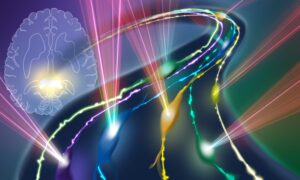
Follow the cellular road
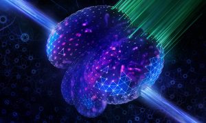
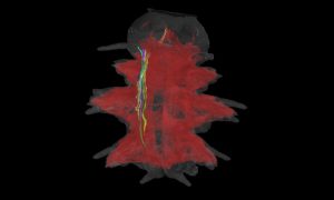
An AI-enhanced advanced microscopy approach offers promise in better understanding glioblastomas, one of the deadliest brain cancers.
SCIENCE & TECHNOLOGY2024
science-technology
Showing results out of
An AI-enhanced advanced microscopy approach offers promise in better understanding glioblastomas, one of the deadliest brain cancers.
SCIENCE & TECHNOLOGY2024
science-technology
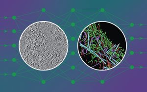
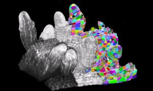
New artificial intelligence tool adds speed and detailed cellular information to analysis of cryo-electron tomography to aid researchers’ understanding of inner cell workings.
2023
science
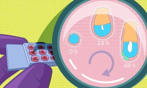
A recent study by EMBL researchers proposes a new method to grow early embryos in the laboratory. With a 3D culture set-up, scientists can closely monitor the changes embryos undergo around the time of implantation.
SCIENCE & TECHNOLOGY2022
sciencescience-technology
EMBL scientists and colleagues have developed an interactive atlas of the entire marine worm Platynereis dumerilii in its larval stage. The PlatyBrowser resource combines high-resolution gene expression data with volume electron microscopy images.
SCIENCE & TECHNOLOGY2021
sciencescience-technology
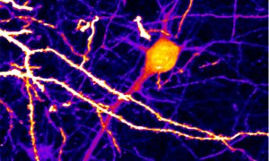
EMBL scientists have combined artificial intelligence (AI) algorithms with two cutting-edge microscopy techniques.
SCIENCE & TECHNOLOGY2021
sciencescience-technology
The Chan Zuckerberg Initiative has recognised four EMBL researchers with their most recent awards, showing how tech trailblazers are integral to advancing science and medicine.
EMBL ANNOUNCEMENTSLAB MATTERS2020
embl-announcementslab-matters
Starting with computer code and moving on to a more user-friendly graphical interface called PlantSeg, the Kreshuk Group at EMBL and collaborators built a simple open-access method to provide the most accurate and versatile analysis of plant tissue development to date.
SCIENCE & TECHNOLOGY2020
sciencescience-technology
Researchers from all life science disciplines – from fundamental biological research to medical applications – generate immense datasets. Analysing these datasets and gaining new knowledge from them is a growing challenge for scientists. The fields of artificial intelligence (AI) and machine…
LAB MATTERSSCIENCE & TECHNOLOGY2020
lab-mattersscience-technology
The image shows a larva of Platynereis dumerilii, a marine worm. The image here was produced by Constantin Pape, a visiting predoctoral fellow in the Kreshuk group at EMBL Heidelberg.
SCIENCE & TECHNOLOGY2020
picture-of-the-weekscience-technology
No results found