Edit

EMBL Taxonomy:

Electron Microscopy Core Facility

Year
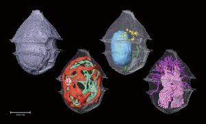
5 October 2023 A composite image of four 3D micrographs showing the cellular organelles of a phytoplankton marked in different colours.

Spotlight: Seeing into the seas

Science & Technology A new method developed by EMBL scientists can help us identify and investigate plankton species in field samples with greater speed, accuracy, and resolution than ever possible before.

2023

sciencescience-technology

5 October 2021 Illustration of a globe with colourful shapes and symbols superimposed.

A cellular atlas of an entire worm

Science & Technology EMBL scientists and colleagues have developed an interactive atlas of the entire marine worm Platynereis dumerilii in its larval stage. The PlatyBrowser resource combines high-resolution gene expression data with volume electron microscopy images.

2021

sciencescience-technology

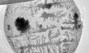
22 September 2020 Black and white electron microscope image of Anopheles mosquito gametes, looking much like feathery fern leaf stencils

A bloom of crystals

Science & Technology How does your crystal garden grow? EMBL's Electron Microscopy Core Facility was able to capture this garden of blooming crystals as they studied mosquito reproductive cells.

2020

picture-of-the-weekscience-technology

EMBLetc.

Looking for past print editions of EMBLetc.? Browse our archive, going back 20 years.

EMBLetc. archive

Newsletter archive

Read past editions of our e-newsletter

For press

Contact the Press Office
Edit