
Spotlight: Colours of the colon
Mucus present in the mouse colon can be visualised using Alcian blue staining, as imaged here by EMBL predoctoral fellow Linda Decker.
Have you recently suffered from a runny nose – whether due to a seasonal allergy or a bout of the flu? Mucus – more colloquially known as snot – is a slimy, watery solution that specialised glands in various parts of our body produce. While mucus may feel like a nuisance that has one always running for a tissue, it actually contains multiple antimicrobial substances and often acts as a layer of protection against pathogens.
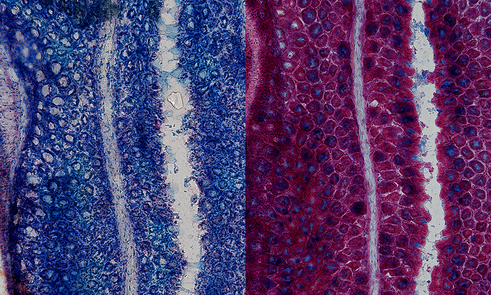
In addition to the respiratory system, our digestive tracts also produce mucus, where it acts as a lubricant, helping food pass through easily and preventing inflammation. In these images taken by Linda Decker, a predoctoral fellow in the Diz-Muñoz Group at EMBL Heidelberg, we can see the mucus layer in the gut stained using Alcian blue, a dye first patented in 1947 as a textile dye and later used by biologists to visualise mucus.
The image shows two cross-sections of the mouse colon, with mucus stained in blue and cell nuclei shown in red. The section on the right has been treated with a mucolytic agent – a chemical that dissolves mucus – bringing down the intensity of the blue staining. In the second image, we can also identify small chambers in the colon, called ‘crypts’, where mucus is produced and secreted.