Welcome: Timo Zimmermann
New team leader looks forward to matching high-tech imagery solutions with biologists’ research needs

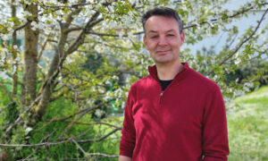
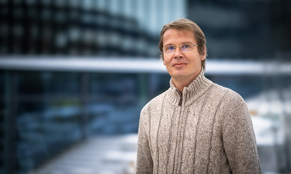
After 13 years helping to nurture and grow the Advanced Light Microscopy Unit at the Centre for Genomic Regulation (CRG) in Barcelona, Timo Zimmermann returns to EMBL, where he cultivated his interest in advanced light microscopy early on. With the new EMBL Imaging Centre set to open in 2021, Timo now has his sights set on the opportunities ahead of him as a team leader who will provide advanced light microscopy services using the latest-generation systems, including technologies developed at EMBL. A biologist by training, Timo is excited by the challenges ahead.
How will being at EMBL take your research to the next level?
In the past few years I’ve mainly been interested in establishing super-resolution imaging methods in my unit and understanding molecular ‘dark states’ that drive super-resolution imaging on the sample side. Here at EMBL, the idea is to offer external researchers timely access to instruments not yet widely available. We will provide the highest-resolution technologies and other advanced methods with a strong focus on connecting to the electron microscopy side of the EMBL Imaging Centre through correlative light and electron microscopy – or CLEM – and cryo-CLEM approaches. In addition, I will create a research team to work on new microscopy techniques in the fields of super-resolution and cryo-fluorescence microscopy.
What makes super-resolution microscopy and cryo-fluorescence microscopy so useful?
Fluorescence imaging under extremely cold, or cryogenic conditions is very useful because it adds context at the light microscopy level that ideally can be combined with the exquisite details visible in cryo-fixed samples in the electron microscope (e.g. cryo-CLEM). But cryo-fluorescence microscopes are still technically limited and very far away from the resolution of electron microscopes. Super-resolution methods could improve the resolution further and allow better correlation, but they require a lot of light to produce the desired improvements at the risk of destroying fine structural details through heating and ice crystal formation. We face interesting challenges to optimally balance all imaging factors to develop cryo-super-resolution workflows that users can employ at the new EMBL Imaging Centre.
What led you to the work you do now?
From the beginning, I focused on microscopy and imaging. I’ve moved from classical electron microscopy to 3D light microscopy to confocal microscopy, which I helped set up in my institute during my PhD. Since I was responsible for the confocal microscope, I started to operate it like a facility, even though there was no official concept then. This helped me apply confocal microscopy to different biological systems and to realise that imaging facilities pose very fulfilling, diverse challenges for me professionally. That led to EMBL, where I enjoyed applying cutting-edge technologies to challenging biological questions. A personal favourite was imaging the transition of malaria parasites through the midgut in live mosquitoes. Working out equations for new spectral imaging approaches helped strengthen my grasp of the mathematics needed in biological imaging, as an interface between complex biological questions and the physics that defines microscopy. This is now central to how I develop imaging approaches.
If you could invite three scientists for coffee, who would they be?
I very much enjoy drinking coffee, so this would be a lively exchange. Because books can provide simple facts, my coffee participants would need to be interesting beyond mere research achievements. Santiago Ramón y Cajal, a Spanish pioneer in neuroanatomy, used the microscope as a tool for discovery and to trace intricate 3D structures with his eye and hand with a camera lucida. He showed how to transform observation into knowledge. This is something we all try to do in our scientific illustrations. Carl Sagan, also, is one of the world’s best science communicators. His TV series Cosmos strongly influenced me growing up. Lastly, movie director James Cameron seems to be an outstanding engineer and out-of-the-box problem solver who pushes boundaries in his creative process. Much of this mindset can also be applied to scientific experimentation.
What has been the biggest challenge in your career so far and how did you overcome it?
I’ve encountered the biggest challenges not in science, but in science politics at the national and European level. This seems especially true if you lead an initiative and need to faithfully represent the diverse interests of all. It requires a sense of balance, a lot of patience, and a very high frustration threshold. I try to look at the whole picture, identify the aim that serves all participants most fairly, and work for this from the beginning. The rest is persistence.
What was your first microscope?
A few weeks into my first biology semester, a lecturer gave me a place in the electron microscopy lab. I remember taking a sample of a snail’s stomach contents to the microscope and finding a weird spiral structure in leaf remnants that I identified as the xylem, a part of a plant’s internal structure for transporting water. Years later, I was at the electron microscope when we studied an unknown 25-million-year-old specimen embedded in amber. Recognising the signature structure of the xylem in that sample brought me back full circle to my first days as a student.
What’s the best part of your work?
Biological imaging is an extremely active field, so it never gets boring. Continuous technical innovations in both light and electron microscopy enable exciting new biological findings. This satisfies both my technological and biological curiosity, and there is no end in sight.


