Spotlight: Using light and sound to see into the brain
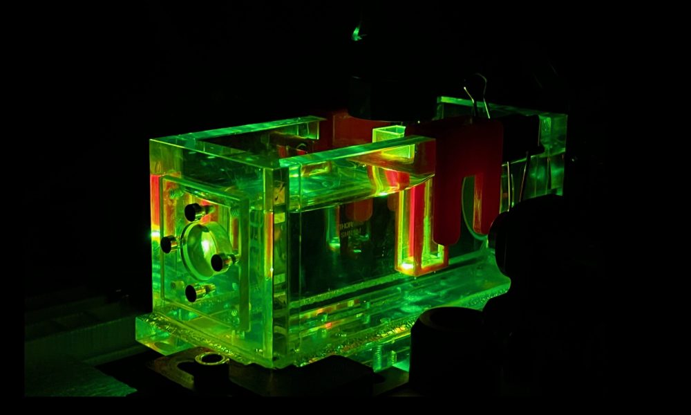
Researchers in the Prevedel Group use this spectroscopy setup to test and optimise photoacoustic probes before their usage in mouse neuroscience.

For biological imaging, scientists have traditionally relied on either light (light microscopy), electron beams (electron microscopy), or sound (sonography). However, by combining light and sound in new and exciting ways, an emerging technique called photoacoustics can generate high quality images deep inside living tissue.
This image, taken by Nikita Kaydanov, predoctoral fellow in the Prevedel lab, shows a pulsed green laser exciting a calcium-sensitive probe made for photoacoustic imaging. This setup was developed in the Prevedel lab and is used extensively in collaboration with the Deo lab.
Photoacoustic spectroscopy provides information about the photoacoustic efficiency of probes, which is a measure of how strongly they generate a photoacoustic signal in the presence of calcium. The custom setup built by the Prevedel Lab measures the photoacoustic signal of specially developed calcium-sensitive molecules in order to steer their development, which is performed by the Deo group at EMBL. These molecules can be used to visualise brain activity with photoacoustic imaging, which allows the scientists to go much deeper into a brain region than other light-based neuroimaging methods.
Ultimately, the Prevedel group aims to image calcium activity throughout the brain of a living mouse, which will provide a new tool for neurobiologists to answer current questions on how the brain functions.


