Scientists collaborate to customise top-of-the-line microscopy method with AI to better understand glioblastoma brain tumours
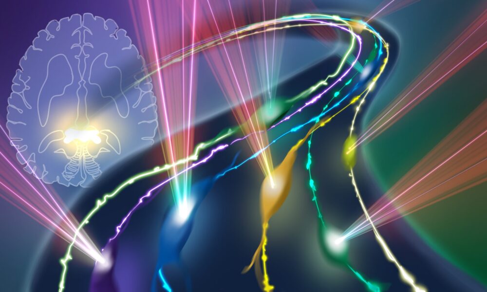
The advanced imaging approach could enable scientists to observe glioblastoma tumour cells within their microenvironment in the white matter – a capability currently lacking and crucial to understanding how tumour cells invade the densely myelinated (insulated) fibre ‘lanes’ of that corpus callosum superhighway, adapting and spreading throughout the brain. Credit: Isabel Romero Calvo/EMBL
Summary
New research provides the proof of concept for a pioneering microscopy technique that can penetrate deep brain tissue.
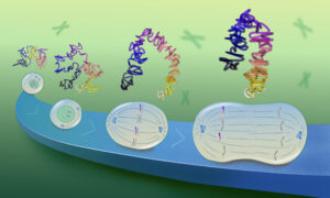
Researchers enhanced the technology with artificial intelligence to enable it to track individual brain tumour cells migrating along a nerve fibre ‘superhighway’ known as the corpus callosum. They specifically targeted cancer cells related to glioblastomas, one of the deadliest brain cancers.
The scientists believe that understanding early cancer cell ‘traffic patterns’ along the corpus callosum could help establish a biomarker for detecting glioblastomas earlier in patients, potentially leading to better diagnostic tools in the future.
Imagine building a traffic surveillance camera that could detect trouble-making cells speeding around in your brain before their cellular gang could commit ‘crimes’. Most importantly, this camera could catch some of the biggest interlopers of all – cancer cells.
This ‘surveillance camera’ is no longer a figment of the imagination. Along the brain’s biggest superhighway of nerve fibres that connects the brain’s right and left hemispheres – the corpus callosum – travel cells that comprise one of the deadliest brain cancers, glioblastomas. Now, scientists have made this cellular detector a reality, introducing artificial intelligence to a state-of-the-art microscope. They can now visualise and track specific cells with unprecedented clarity in deep brain tissue, including along this superhighway.
In a recent collaborative endeavour between EMBL and Heidelberg University, scientists are using this new technology to track glioblastoma tumour cells to better understand this deadly cancer and possibly detect it earlier, which could potentially lead to better diagnostic tools in the future.
A deep tissue microscope is born
In 2021, EMBL researchers – with collaborators from Germany, Austria, Argentina, China, France, the United States, India, and Jordan – developed a new microscopy technique. EMBL Group Leader Robert Prevedel and his research group worked with these diverse collaborators to address some of the challenges neuroscientists face in studying deep brain regions. Previously, diffuse brain tissue posed a problem for scientists when they tried to observe neurons and glial cells known as astrocytes and study how they communicate deep within the cortex. It also made it difficult to visualise neural cells in the hippocampus, another deep brain region responsible for spatial memory and navigation.
The scientists based their new approach on state-of-the-art microscopy methods that could provide a wider and clearer aperture for viewing while also adjusting for the distortion that arises when light waves scatter in deep brain tissue. They envisioned many possible future applications in brain research.
Now, in a study published in the journal Nature Communications, Prevedel has teamed up with neuroscientists, neurooncologists, and AI experts to take this microscope to the next level. The result is a microscope that can observe living neurons – and other kinds of brain cells – deep within the brain over a prolonged period of time.
“We have now gone from taking a snapshot of cells in a mouse brain to zooming in on specific cells and being able to follow them for many hours or even days,” Prevedel said. “Also, incorporating custom AI approaches has allowed us to distinguish different parts of the microenvironment of the cells, which is also very important to understand their behaviour in context.”
Putting it to the test
In 2021, Varun Venkataramani at the Neurology Clinic of the University Hospital Heidelberg read of this new approach to deep-tissue microscopy with great interest. His research focuses on human brain tumours – notably glioblastomas, which are prevalent, fast-growing, and intractable tumours. Venkataramani was learning more and more about neural mechanisms that determine how tumours originate, progress, and ultimately respond to or resist treatment. However, his microscopy approach at the time restricted imaging depth, limiting them mainly to the brain’s grey matter.
“The 2021 paper by Robert’s group introduced a deep-tissue microscopy technique that I believed could extend our imaging capabilities to the white matter of the corpus callosum,” Venkataramani said. White matter plays a role in communication between different grey matter areas of the brain and the rest of the body. “This could potentially reveal novel biological processes and offer insights into the behaviour of these tumours in a critical, yet understudied niche,” he added.
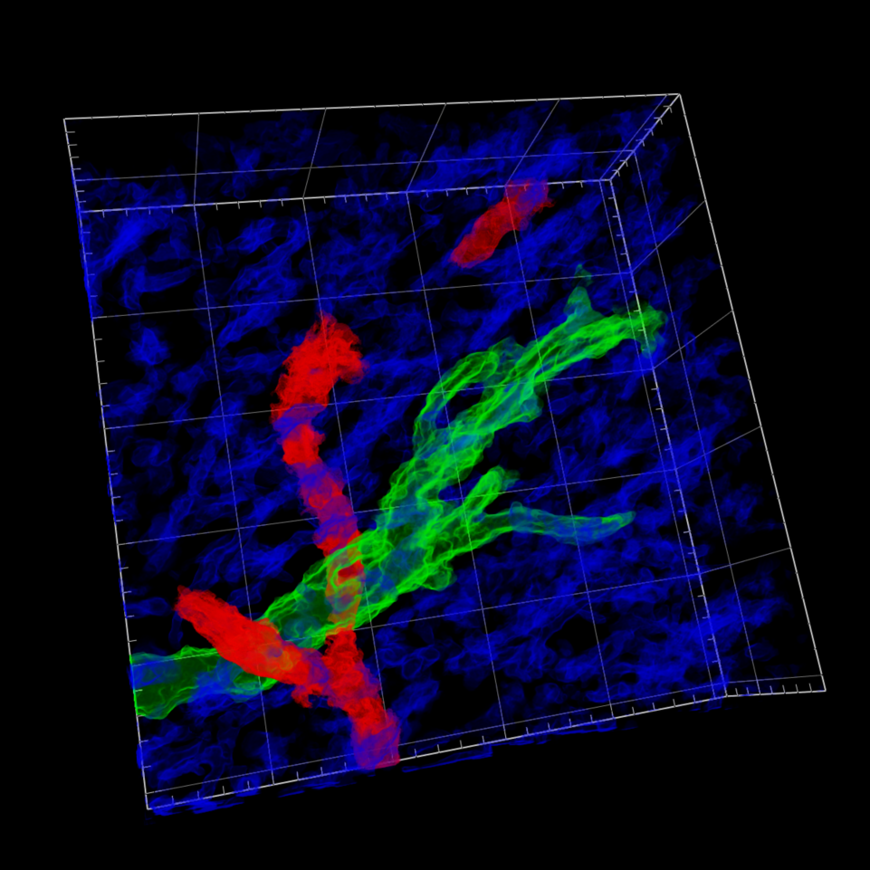
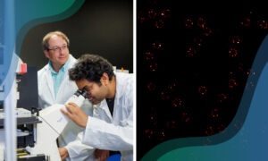
A glioblastoma tumour cell (green) present in the white matter (blue) near a blood vessel (red), visualised via the novel three-photon microscopy workflow Deep3P. Credits: Stella Soyka/Universitätsklinikum Heidelberg, Marc Schubert/Universitätsklinikum Heidelberg, Robert Prevedel/EMBL, Varun Venkataramani/Universitätsklinikum Heidelberg.
Glioblastomas are primarily a white-matter disease. The new advanced imaging technique enabled Venkataramani’s team to observe these tumour cells within their microenvironment in the white matter. This capability was crucial to understanding how tumour cells invade the densely myelinated (insulated) fibre ‘lanes’ of the corpus callosum superhighway and then adapt and spread throughout the brain. This process is also associated with glioblastomas’ lethally invading critical brain structures.
“It has been fascinating to observe tumour cell invasion in the corpus callosum in real-time,” said Marc Schubert, one of the study’s lead authors and medical student at Heidelberg University.
“At this point, I think the most important aspect of this fundamental research is that it allows us to investigate these tumours in their most relevant microenvironmental niche for the first time,” Venkataramani said. “These findings also help explain the current challenges in detecting glioblastoma cells at the tumour’s infiltrative edges using conventional MRI techniques, which are the standard in clinical imaging. As a neuroscientist, neurologist, and neurooncologist, I see potential for this technology to bridge the gap between laboratory research and clinical application, improving how we could diagnose and potentially treat brain tumours.”
Artificial intelligence takes microscope to the next level
An important feature of this latest collaboration was that the researchers incorporated an element of artificial intelligence.
“From a technical development point of view, the AI-based methods helped to ‘denoise’ our images, so the contrast now is much clearer,” Prevedel said. “The AI can distinguish different structures inside the white matter, like myelinated fibres and blood vessels, which is important for a variety of reasons. AI was really instrumental in advancing the level of this microscope, so it can address these pressing medical questions.”
Anna Kreshuk’s research group at EMBL Heidelberg provided this AI expertise. Kreshuk’s group contributed to a customised workflow that helped distinguish blood vessel signals from the myelinated neural fibre ones, clarifying the tumour cells’ microenvironment.
Consequently, the researchers could identify a potential microscopic imaging biomarker linked to the structural properties of the white matter microenvironment. This innovative workflow sets the stage for potentially identifying imaging patterns for glioblastomas, so tumours could be detected earlier than they are currently.
“We are looking forward to further customising this new approach towards more clinically practical needs in the future to maximise its potential,” said Stella Soyka, one of the paper’s lead authors and a medical resident in the Department of Neurology at Heidelberg University Clinic.
“It’s promising, but it’s much too soon to apply it clinically without further development,” Venkataramani said, explaining that the next steps will integrate further advanced imaging modalities, which can help build practical tools for standard clinical settings.
“We are optimistic, particularly because of the robust interdisciplinary support from the Heidelberg-Mannheim Life Science Alliance network, which fosters collaboration across preclinical and clinical disciplines,” he said. “This synergy is crucial for bringing these laboratory insights into clinical practice in the foreseeable future.”