Actin moves chromosomes
Discovery changes fundamental thinking
Microtubules need a helping hand to find chromosomes in dividing egg cells, scientists have discovered. Although it was generally accepted that microtubules act alone as the cellular ropes to pull chromosomes into place, a new study by researchers at the European Molecular Biology Laboratory (EMBL) shows that this is not the case. They found that in large cells such as animal eggs, something else is needed to move the chromosomes into the correct location – fibres of the cytoskeletal molecule actin (Nature, 13 July 2005)
“No one has ever shown that actin moves chromosomes,” says Dr. Jan Ellenberg, the EMBL researcher whose group carried out the research. “We were able to do so because our group is one of the few that studies cell division in starfish – an ideal model for observing division in living animal eggs.”
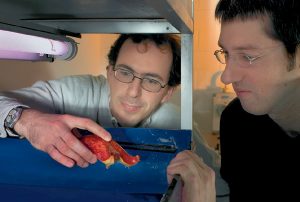
The starfish is an excellent model for studying oocytes, the cells that give rise to egg cells. In this marine animal, these cells are transparent and mature quickly outside the body, and can be kept alive in a drop of seawater. That’s why EMBL scientists performed some of their experiments with collaborators at the Marine Biological Laboratory in Woods Hole, MA, USA – working with animals fresh from the ocean.
Ellenberg and PhD student Péter Lénárt studied the starfish oocytes as they underwent meiosis, a special cell division that is needed to halve the number of chromosomes in an egg before it unites with a sperm. When the protective nuclear membrane surrounding the chromosomes breaks down during meiosis, it was thought that microtubules capture the chromosomes and act as ropes to pull them to the surface and expel half of them from the cell.
But when the EMBL researchers measured the microtubules, they discovered that they were, in fact, much too short to transport the chromosomes over the long distance to the surface of the large oocyte. By using a chemical to disable the microtubules, they found that cells were still able to pull chromosomes into the proper positions.
So what was moving the chromosomes?
When they repeated the experiment with a chemical that breaks down the other major type of cellular fibres, actin, the cells lost track of their chromosomes and the new cells had unequal amounts of genetic material. This condition, called aneuploidy, is thought to be a major cause of miscarriages and some types of birth defects.
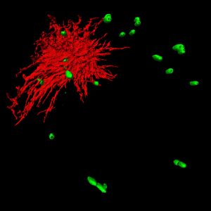
Lénárt spent 18 months optimizing an imaging technology, with help from collaborators at the German Cancer Research Center (DKFZ), to visualize the delicate actin fibres before he could confirm the group’s fundamental breakthrough. He observed a network of filamentous actin forming in the region where the nuclear membrane breaks down. This network acts as a fishnet to gather all the chromosomes together and drag them close to the short microtubules. Only then, when the chromosomes are close enough, can the microtubules latch on and pull half of them outside the cell.
The implications for this pioneering work are clear. Starfish oocytes have many similarities to those of other animals, including humans. Because this mechanism is essential to prevent chromosome loss before fertilization, advances in this field could help to explain the causes of pregnancy loss and birth defects in humans.
