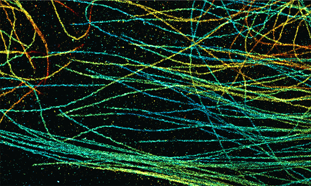
Deep learning enables fast and dense single-molecule localization with high accuracy
Nature Methods 03 September 2021
10.1038/s41592-021-01236-x
EMBL scientists and their collaborators have developed DECODE, a neural network based fitter that accelerates super-resolution microscopy imaging

Scientists use super-resolution microscopy to study previously undiscovered cellular worlds, revealing nanometre-scale details inside cells. The method revolutionised light microscopy and earned its inventors the 2014 Nobel Prize in Chemistry.
Single-molecule localisation microscopy (SMLM) is a type of super-resolution microscopy. It involves labelling proteins of interest with fluorescent molecules and using light to activate only a few molecules at a time. Using this method, multiple images of the same sample are acquired. To create a meaningful picture, a computer program unscrambles the data and compiles the complete image.
While the technique can be used to locate molecules with high precision, it requires scientists to acquire a large number of images. “One of the biggest limitations of super-resolution microscopy is how long it takes to collect data from the microscope,” explained Lucas-Raphael Müller, a former master’s degree student in the Ries Group at EMBL Heidelberg.
In a collaborative effort, the Ries Group, together with the labs of Professor Jakob Macke at the University of Tübingen (Cluster of Excellence – Machine Learning for Science) and Dr Srinivas Turaga at Janelia Research Campus, developed a new algorithm that overcomes several limitations of SMLM.
DECODE (DEep COntext DEpendent) is a computer program based on a neural network that learns from training data and gets better over time as it is fed more and more examples. The scientists used a trick that was recently established for training neural networks: instead of using real images to train the network, they used synthetic data generated by a numerical simulation. By incorporating information about the microscopic setup and the imaging physics, the researchers achieved simulations that closely matched real-world acquisitions. As a result, a neural network that is trained to localise fluorescence molecules using simulated data can also localise fluorophores in real images.
One of the main benefits of DECODE is that it accurately detects and localises fluorophores at higher densities than were previously possible. This means that researchers can activate more fluorophores for imaging in a single camera frame, and fewer images are needed per sample. As a result, imaging speeds can be increased up to tenfold with minimal loss of resolution.
“Instead of looking at frames in isolation, like previous computer programs for super-resolution microscopes, DECODE uses information from the neighbouring frames to get more context,” explained Artur Speiser, a PhD student in the Macke and Turaga labs. In a single image, several fluorescing spots that are close together might look like one molecule. But looking at previous and next images of the same area can reveal that parts of this fluorescent glow switch off at different times, which suggests that they are distinct entities.
“This novel approach is more efficient for imaging activity in living cells, and for studying fragile samples that can break down under extended exposure to harsh light,” said Dr Turaga. “And it is possible to upgrade existing analysis software using DECODE, which will allow many labs performing SMLM to improve their reconstructions, even on existing datasets.”
The artificial neural network DECODE software thus solves several limitations of SMLM. “These research results could be achieved only by the excellent collaboration between the different groups with interdisciplinary backgrounds,” added EMBL Group Leader Jonas Ries. “The software is simple to install and free to use, so we hope it will be useful for many scientists in the future.”
Nature Methods 03 September 2021
10.1038/s41592-021-01236-x