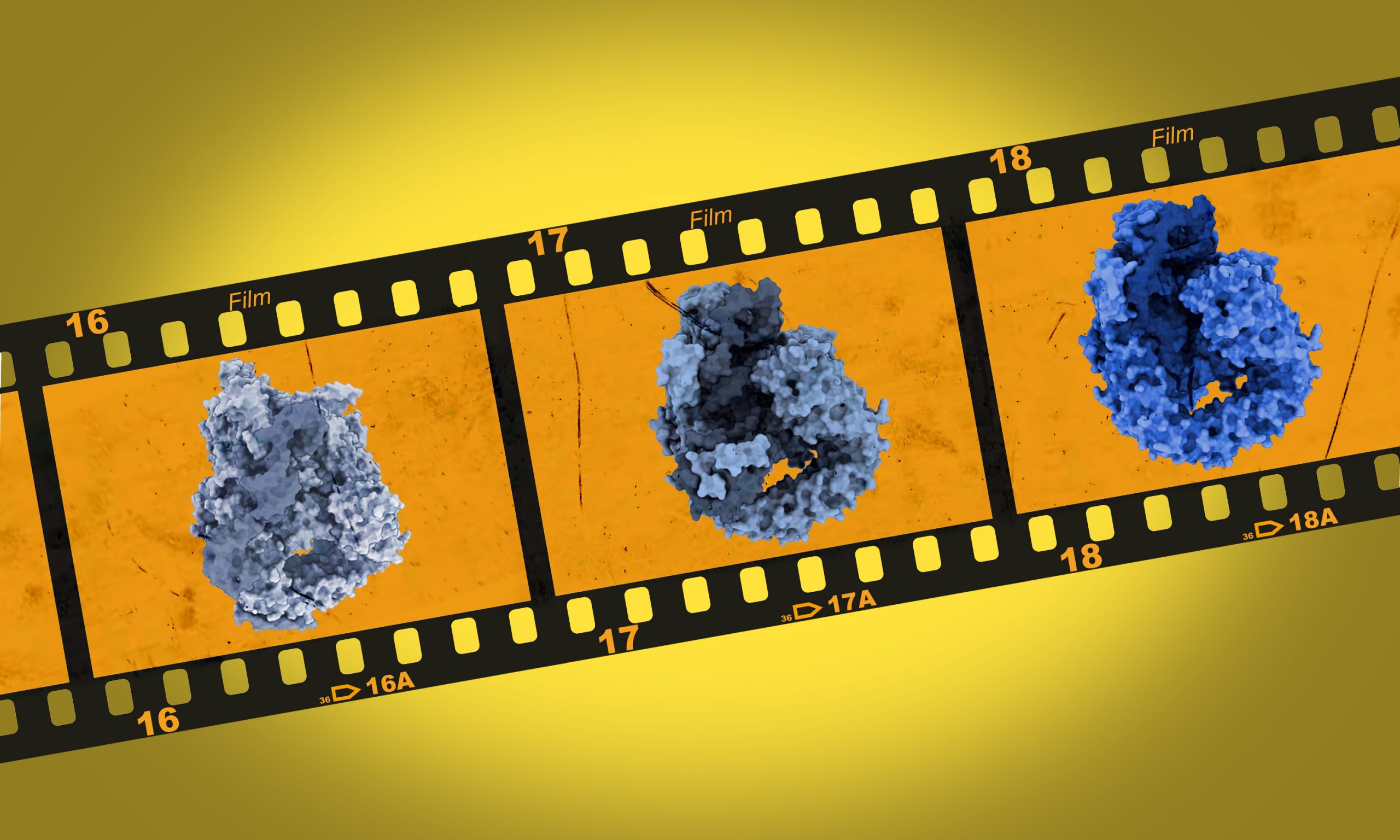
Structural basis of branch site recognition by the human spliceosome.
Science 25 November 2021
10.1126/science.abm4245
Cryo-electron microscopy sheds light on RNA splicing

The Galej group at EMBL Grenoble has recently obtained high resolution snapshots of the dynamic rearrangements of the U2 snRNP complex, a crucial component of the human spliceosome. This macromolecular complex is responsible for sorting out coding from non-coding segments during RNA splicing – the process by which information in messenger RNAs is rearranged, allowing them to produce functional proteins. These results have been published in Science.
DNA makes RNA makes protein. This seemingly simple phrase, describing how genes are transcribed into messenger RNAs and subsequently translated into proteins, hides a multitude of intricate molecular steps. In eukaryotes – organisms like animals, plants and fungi – one of these steps is called RNA splicing.
Splicing is necessary because nearly all genes in eukaryotic cells include segments called introns, which are not required to make a protein. In order to create proteins that function properly, the cell needs to remove introns from the mRNA molecule, retaining only exons – the segments that carry protein-coding information. This reaction is catalysed by a large macromolecular complex called the ‘spliceosome’.
Spliceosomes are complex and highly dynamic molecular machines, which makes them very difficult to study with traditional structural biology methods. Now, cryo-electron microscopy (cryo-EM) techniques have allowed researchers in the Galej group at EMBL Grenoble to show how the spliceosome recognises the branch site, a key position within introns which influences the outcome of the splicing reaction.
Branch sites are located close to the ends of introns. Because of this location, they are important for determining where an intron ends and the protein-coding sequence begins again. This means that without them, a messenger RNA that describes a fully functional protein cannot be produced.
The researchers used cryo-EM to obtain snapshots of the molecular structure of the U2 snRNP complex, the component of the spliceosome that recognises the branch site. “We developed a novel method to purify these complex particles directly from human cells and determined their 3D structure at a very high resolution,” said Jonas Tholen, first author of the paper and PhD student in the Galej group. “Atomic details allow us to see precisely how different proteins and RNAs interact with each other and with the branch site.”
Combining these structures with biochemical assays, the scientists were able to describe in detail the mechanism by which the spliceosome recognises branch sites and executes the splicing process at precisely these locations.
They could find two previously unknown intermediates of the complex during the splicing process and show that an additional protein – SF3B6 – plays an important role in stabilising the branch site within the spliceosome. “A series of biochemical studies from 25 years ago indicated that this protein is very important for the splicing reaction, but its precise function remained elusive. With our new insights, we could explain those early observations,” explained Wojciech Galej.
“Some estimates predict that up to 60% of genetic disorders occur in relation to problems with splicing signals or malfunctions of the splicing machinery,” added Galej. “This makes the splicing machinery a potential drug target for treating some genetic diseases. Understanding the molecular and atomic details of the splicing mechanism opens up new possibilities for identifying or developing drugs that can modulate this process in treating certain genetic diseases.”
Science 25 November 2021
10.1126/science.abm4245