Digital zebrafish embryo provides the first complete developmental blueprint of a vertebrate
New Google Earth™-like model allows zooming in on the development of zebrafish

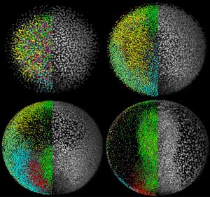
Researchers at the European Molecular Biology Laboratory (EMBL) have generated a digital zebrafish embryo – the first complete developmental blueprint of a vertebrate. With a newly developed microscope scientists could for the first time track all cells for the first 24 hours in the life of a zebrafish. The data was reconstructed into a three-dimensional, digital representation of the embryo. The study, published in the current online issue of Science, grants many new insights into embryonic development. Movies of the digital embryo and the underlying database of millions of cell positions, divisions and tracks will be made publicly available to provide a novel resource for research and scientific training.
To get from one cell to a complex organism, cells have to divide, travel around the body and arrange intricate shapes and specialised tissues. The best way to understand these dynamic processes is to look at what happens in the first few hours of life in every part of an embryo. While this is possible with invertebrates with a few hundred cells, like worms for example, it has so far been impossible to achieve for vertebrates.
“Imagine following all inhabitants of a town over the course of one day using a telescope in space. This comes close to tracking the 10 thousands of cells that make up a vertebrate embryo – only that the cells move in three dimensions,” says Philipp Keller. Together with Annette Schmidt he carried out the research in the labs of Jochen Wittbrodt and Ernst Stelzer at EMBL.
Two newly developed technologies were key to the scientists’ interdisciplinary approach to tracking a living zebrafish embryo from the single cell stage to 20,000 cells: a Digital Scanned Laser Light Sheet Microscope that scans a living organism with a sheet of light along many different directions so that the computer can assemble a complete 3D image, and a large-scale computing pipeline operated at the Karlsruhe Institute of Technology.
Zebrafish is a widely used model organism that shares many features with higher vertebrates. Taking more than 400,000 images per embryo, the interdisciplinary team generated terabytes of data on cell positions, movements and divisions that were reassembled into a digital 3D representation of the complete developing embryo.
“The digital embryo is like Google Earth™ for embryonic development. It gives an overview of everything that happens in the first 24 hours and allows you to zoom in on all cellular and even subcellular details,” says Jochen Wittbrodt, who has recently moved from EMBL to the University of Heidelberg and the Karlsruhe Institute of Technology.
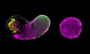
New insights provided by the digital embryo include: fundamental cell movements that later on form the heart and other organs are different than previously thought and the position of the headtail body axes of the zebrafish is induced early on by signals deposited in the egg by the mother.
The new microscopy technology is also applicable to mice, chickens and frogs. A comparison of digital embryos of these species is likely to provide crucial insights into basic developmental principles and their conservation during evolution.
Download the movies from www.embl.de/digitalembryo


