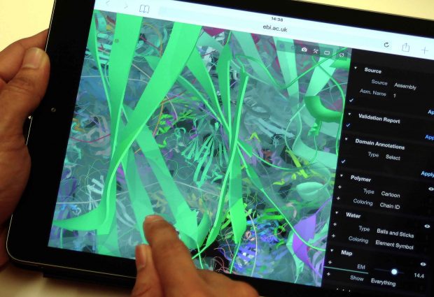
Exploring 3D macromolecules in a flash
New techniques from gaming allow users to load giant 3D protein images quickly in PDBe

EMBL-EBI’s Protein Data Bank in Europe (PDBe) has updated its 3D viewer, enabling users to load 3D protein structures instantly on any Internet-connected device. The upgrade particularly benefits researchers working in areas with low Internet speed. The new viewer is also mobile-friendly, allowing anyone who is interested in biology to explore the beauty and mystery of proteins and other macromolecules.
PDBe prides itself on being the European resource for collecting, organising and sharing data on molecular structures, such as proteins. These ‘macromolecules’ include proteins and nucleic acids, plus carbohydrates, lipids and drug molecules represented in more than 130,000 PDB entries. The team also develops free analytical tools to make structure-related data more and more accessible to the biomedical community.
Macro molecules, massive files
Some of PDBe’s 3D structures, are huge, notably those solved by electron microscopy, for which the Nobel Prize was awarded recently. Nevertheless, each detail is visible and easy to explore. Delivering this amount of detail comes with a price. Files can exceed a gigabyte, so they load very slowly, especially on older machines or in areas with poor Internet connectivity. This also makes the structures hard to view on devices such as mobile phones or tablets.
To tackle this challenge, the PDBe team, working closely with scientists from the Central European Institute of Technology (CEITEC) in the Czech Republic, has implemented a compression format that reduces the size of files up to 500 times. The new viewer, LiteMol Suite, also uses a technique employed in virtual reality and gaming, which involves downloading only essential data, such as one section of a protein, or a small part of the experimental data, instead of the whole file.
A world of wonder
“Seeing the intricate patterns of a virus or visualising how a pain-killing drug works are unique experiences that have traditionally needed specialised software,” explains Matthew Conroy, Scientific Curator at PDBe. “Public databases and visualisation tools are becoming much more accessible, which means the biological wonders we work with are open to a much wider audience.”
The PDBe team hopes that the implementation of LiteMol will encourage more people – including teachers and pupils – to explore EMBL-EBI’s free structural data resources.
“In the UK, students as young as 13 are studying proteins as part of the science curriculum,” continues Conroy. “PDBe’s new viewer could support classroom teaching, giving students a chance to actually see a protein’s structure in 3D, play around with it, zoom in and explore.”
For a more detailed look at PDBe’s interactive, web-based visualisation of large-scale macromolecular structure data, read the team’s recent paper in Nature Methods.
This post was originally published on EMBL-EBI News.


