MOrgAna: accessible quantitative analysis of organoids with machine learning
Development 8 September 2021
10.1242/dev.199611
EMBL scientists created an open-source, user-friendly platform specialising in morphology and fluorescence
Within the space of a few days in March 2020, Nicola Gritti and Jia Le Lim from the Trivedi Group at EMBL Barcelona had to stop their experiments and stay home. In response to the pandemic, EMBL had restricted who could work on-site. Not knowing when it would be possible to restart their lab activity posed a challenge for the research group. “We decided to start analysing the huge amount of images we had from our previous experiments, and needed a tool to segment and easily share the results with each other,” said Gritti. The results of these efforts have now been published in Development.
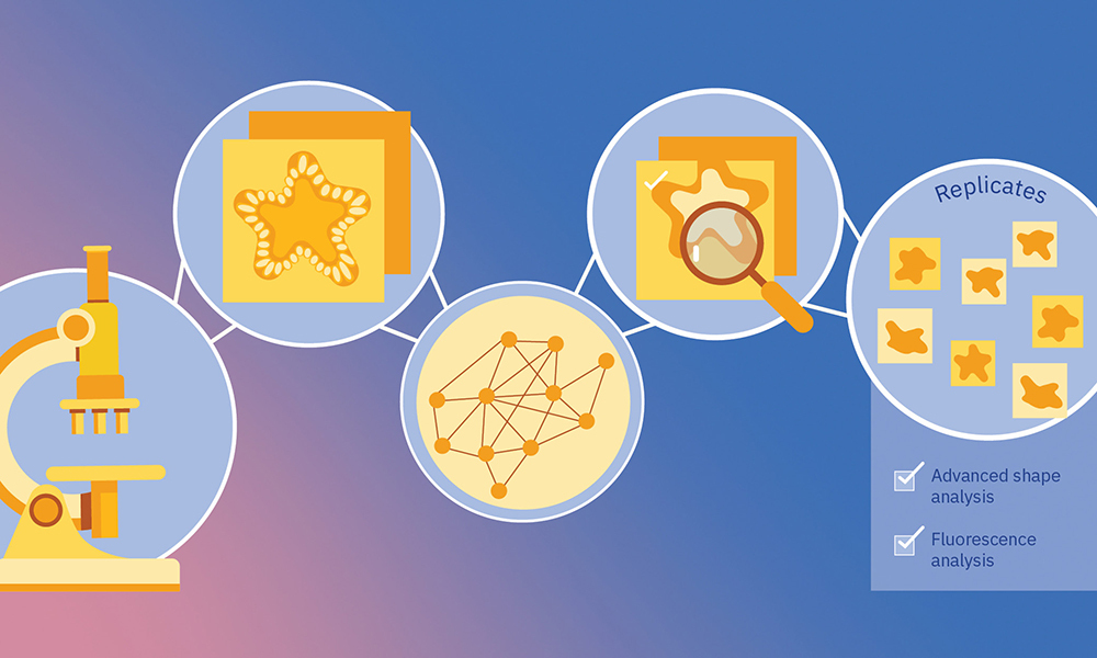
Within a year, the researchers had developed MOrgAna, open-source software that uses machine learning to analyse images of organoids and perform segmentation – the process of extracting cellular components from an image. MOrgAna (1) has a user-friendly modular design and requires no programming experience to use, (2) can also be customised and integrated into advanced image analysis pipelines, (3) and can quantify morphological features.
“The priority was to build a user-friendly platform able to analyse hundreds of organoids and have a graphical overview of their biological results,” said Lim. The software can analyse images of several types of samples – human brain organoids, zebrafish explants, and intestinal organoids, among others – that have been acquired with a range of microscopy devices, from stereoscopic microscopes to confocal or high-content screening microscopes.
MOrgAna is not just a pretty face; it also performs better segmentation than existing platforms due to its machine-learning algorithms. Such algorithms are capable of learning non-linear relationships between the input images and the ground truth. More concretely, MOrgAna learns to classify the pixels in an image into three types: background, organoid, and organoid edge. This is particularly important, as organoids develop over several days; dying cells surround the edge of the organoids, making it difficult to detect their boundaries accurately.
Upon segmentation, MOrgAna can be used to quantify several morphological and fluorescence features. Thanks to the platform’s modular format, quantification can be adapted to each experiment, organoid type, and relevant features. Whereas most platforms quantify simple parameters like area, perimeter, and average fluorescence intensity, MOrgAna captures complex morphological features of organoids, such as the number of protrusions, using LOCO-EFA (lobe contribution elliptic Fourier analysis). In addition, the platform can identify the axes of organoids to quantify fluorescence intensity along the primary (longest) axes, as well as their angular and radial profiles.
MOrgAna overcomes the limitations of traditional tools, which often cannot distinguish between normal conditions and the effects of mutation, infection or drug treatment. And by being freely accessible, the platform is available for use by scientists around the world. Both Gritti and Lim highlighted the importance of open-source technologies for scientific advancement: “Platforms like MOrgAna enhance the natural interaction between scientists of different specialities, which can push existing research and foster new interdisciplinary fields.”
MOrgAna is a clear example of the present and future of science: open, collaborative, and designed to speed up knowledge transfer and tackle societal challenges without delay.
Development 8 September 2021
10.1242/dev.199611
