Shining high-brilliance beams on coronavirus structure
EMBL researchers are using small-angle X-ray scattering to study the structure and interaction of SARS-CoV-2 molecules

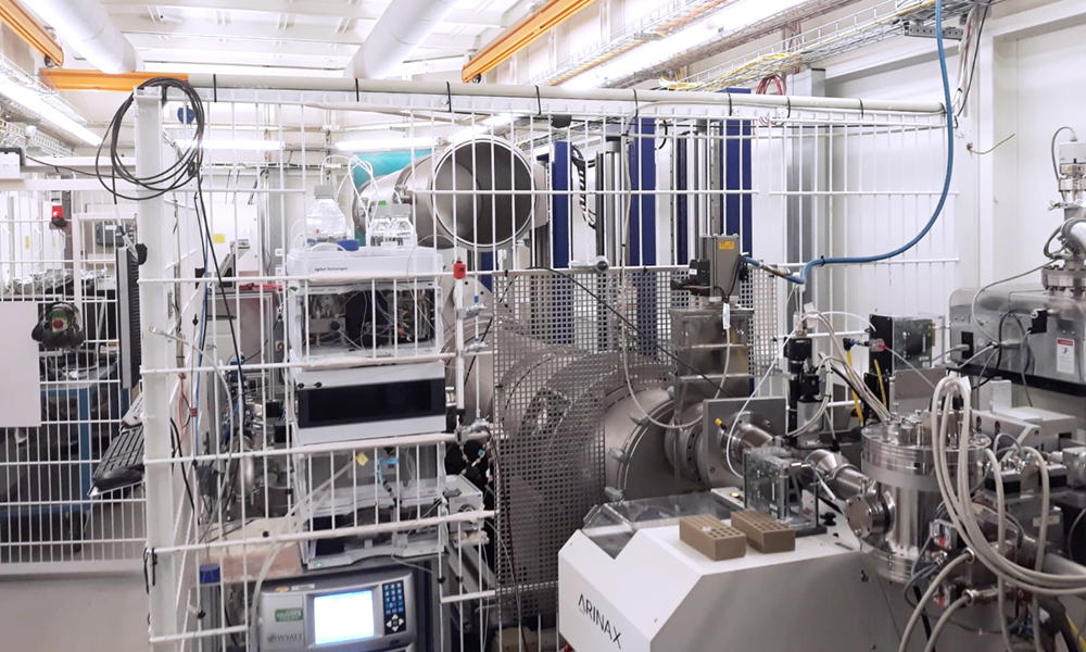
EMBL researchers are studying COVID-19-related molecules by exposing them to high-brilliance X-ray beams. The Svergun group at EMBL Hamburg is using biological small-angle X-ray scattering (SAXS) as part of a global effort by scientists to elucidate the structural organisation of SARS-CoV-2 proteins, identify key antibodies, and pinpoint molecular drug targets to hopefully halt the virus in its tracks.
SAXS makes it possible to reconstruct the 3D shapes of crucial molecular units in a cell or virus. Intense X-ray radiation, produced using the PETRA III synchrotron, is fired at proteins in solution. The radiation that scatters off the protein molecules is collected and combined with modelling and computation. Experiments can be carried out in seconds, and the first models of molecular structure are available within minutes. It is possible to observe changes to proteins over time, in conditions comparable to those inside a living cell. Alone, or in combination with X-ray crystallography, nuclear magnetic resonance, or electron microscopy, SAXS provides a powerful way of pinpointing the roles of cellular and viral machinery during infection.
In one study, the EMBL researchers are using SAXS to rapidly screen neutralising antibodies at high speed and identify those binding to SARS-CoV-2’s distinctive spike proteins, which the virus uses to enter the cell and initiate infection. Another study aims to find small molecular compounds that can disrupt the coronavirus replication machinery by preventing the dimerisation of an essential viral protease thought to be a prime target for inhibitors. The group is also collaborating with other researchers who support the further use of SAXS in coronavirus-related experiments. The work could contribute towards the validation and development of tests and treatments for COVID-19.


