Optogenetic control of apical constriction induces synthetic morphogenesis in mammalian tissues
Nature Communications 14 September 2022
https://doi.org/10.1038/s41467-022-33115-0
EMBL Barcelona researchers have developed a tool that can use light to control the shape of cells

Being able to change the shape of cells in the lab gives researchers a way to mimic morphogenesis, the biological process by which cells organise to form 3D tissues and organs. Researchers in the Ebisuya Group at EMBL Barcelona have developed a novel optogenetic tool, which allows them to control cell and tissue shape by shining light on them. Their work was recently published in Nature Communications.
During embryonic development, cells multiply, move, and come together to form different kinds of tissues, eventually creating a whole organism.
“Our lab studies synthetic developmental biology, which is a branch of science that builds tissues and tries to control them to understand how they function,” said Guillermo Martínez-Ara, first author of the paper, who recently completed his PhD in the Ebisuya group. “In this particular project, we were interested in looking at how cells change shape to form tissues.”
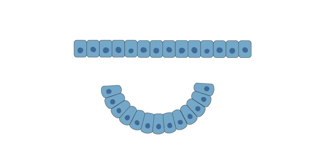
Apical constriction, a process by which a cell actively reduces its top surface, is necessary for the formation of numerous curved structures in embryos. If we imagine a cube-shaped cell, when it decreases the area of its top surface – the apical part – it becomes pyramidal. When a row of such cube-shaped cells sit next to each other and all undergo apical constriction at the same time, the row changes shape from a straight line to a U-shaped one. Apical constriction thus helps organisms form curved tissues during early development.

Ebisuya’s team modified a protein that triggers apical constriction – called Shroom3 – to make it sensitive to light. This modified version of the protein, which the team calls OptoShroom3, can be activated or deactivated by switching blue light on and off. Researchers observed that upon activation of OptoShroom3, it takes less than a minute for some cell types, like epithelial cells and neural tissues, to undergo apical constriction.
“Apical constriction is crucial for several processes in vertebrates, such as the formation of the gut, the kidney, or the early stages of the optic cup in the eye,” said Martínez-Ara. “Our modified version of shroom3 can rapidly activate apical constriction. When we activate OptoShroom3 with light, we can cause tissues to fold, thicken, or flatten.”
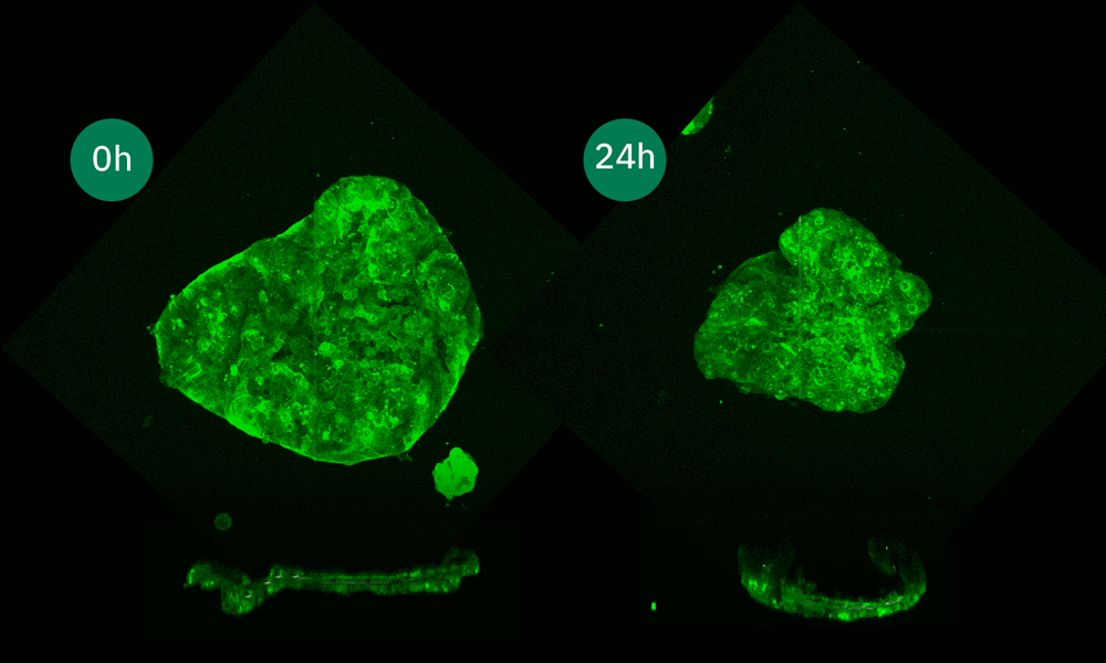
The researchers observed that when they stimulated groups of cells using this method, they could promote cell elongation and collective movement. For example, in colonies of epithelial cells grown on soft gels, stimulation with light induced irreversible folding of cell sheets. In organoids – tiny in vitro tissues resembling organs in their structure and function – apical constriction also altered tissue structure in different ways, proving that OptoShroom3 can be applied to more complex settings.
“Our long-term plan is to use this tool to modulate the shape of organoids.” said Miki Ebisuya, Group Leader at EMBL Barcelona. “Organoids are complex structures and to us, they stand out as perfect candidates to study the interaction between tissue shape and function.”
Nature Communications 14 September 2022
https://doi.org/10.1038/s41467-022-33115-0