Conformational changes in Lassa virus L protein associated with promoted binding and RNA synthesis activity
Nature Communications 2 December 2021
10.1038/s41467-021-27305-5
EMBL structural biology is part of an international collaboration addressing zoonotic disease caused by Lassa virus

Endemic in Western African countries, Lassa virus is transmitted to humans through food or household items that are contaminated with the urine or faeces of Mastomys rats. Even though many people who become infected with Lassa virus are asymptomatic, one in five infections results in severe haemorrhagic disease, attacking vital organs like the liver, spleen, and kidneys.
The World Health Organization (WHO) lists Lassa fever as a significant public health threat with high epidemic potential and no effective countermeasures. Lassa fever has no vaccine, and only one drug – a broad-spectrum antiviral called ribavirin, which has limited efficacy.
Even though researchers are working on a vaccine, an effective antiviral is needed to reduce the number of severe cases and deaths. This is where structural biology can help.
To understand the intricate mechanism of viral replication, structural biologists focus on a key component: the viral polymerase. In an infected cell, this enzyme replicates the virus’s genetic information. The genome copies are then packaged into new virions, which escape the cell and can infect a new host. Determining the polymerase structure helps researchers to understand how a virus replicates, giving them important insight into how to design drugs that can stop the infection.
Stephen Cusack, head of EMBL Grenoble, focuses his group’s research on the polymerase of several human pathogenic viruses, particularly influenza virus. Because influenza and Lassa viruses are from the same family – both segmented negative-strand RNA viruses – Cusack’s group started collaborating on Lassa virus several years ago with Maria Rosenthal and Stephan Günther from the Bernhard Nocht Institute for Tropical Medicine (BNITM) in Hamburg.
“Stephen Cusack was at the forefront of research on influenza and ahead of the field with respect to bunyaviruses and arenaviruses,” said Günther, the head of the virology department at BNITM. “We thought it was excellent synergy because he already had a lot of technological knowledge we could transfer to the Lassa polymerase.”
The Lassa polymerase has characteristics that complicate the process of determining its structure, which Rosenthal, a BNITM structural biologist and group leader, had tried to obtain for several years. “After a long testing of other systems, I went to Grenoble to test the system developed by Imre Berger (an EMBL Grenoble alumnus), and applied by the Cusack group to influenza. We got very good expression of the protein,” Rosenthal said.
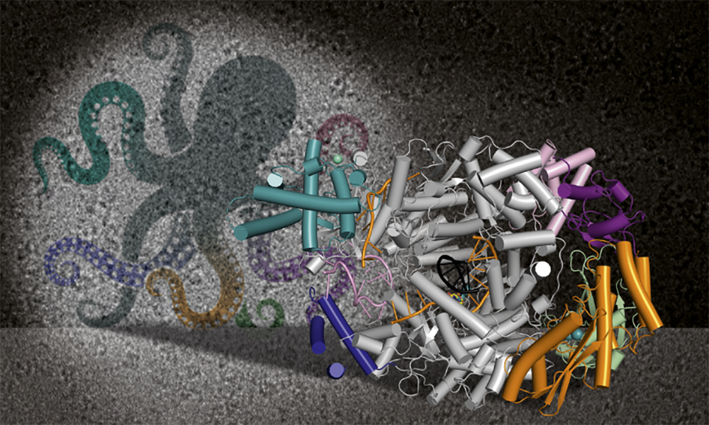
The polymerase also has a peculiar architecture with a core and flexible outer components. “We call the Lassa virus polymerase an ‘octopus’ because it’s like it has arms floating around, which makes it very hard to catch in one position,” said Tomas Kouba, a postdoc in the Cusack Group.
Macromolecular crystallography – an imaging technique commonly used in structural biology – couldn’t be used to observe the Lassa polymerase, because the protein moved too much to capture all the arms. Cryo-electron microscopy (cryo-EM) offered the solution as it enables snapshots of the protein to be taken in different functional states.
“We applied to Lassa the techniques that Kouba and Joanna Wandzik (a former EMBL PhD student) developed to visualise influenza polymerase in action,” Cusack explained. ” We used cryo-EM facilities both in Grenoble and at the Centre for Structural Systems Biology [CSSB] at the University of Hamburg.”
This collaboration between researchers from EMBL Grenoble, BNITM, and Kay Grünewald’s lab at the CSSB resulted in a paper recently published in Nature Communications. It details nine structures of the Lassa viral polymerase in different functional configurations.
“When you observe this ‘octopus’, you also want to see how it behaves, how it moves its arms to eat or catch something,” explained Kouba, one of the paper’s three principal authors.
“The power of cryo-EM is that you take millions of pictures of the protein. Then you can sort them into several boxes: one box with an arm up, and then one box with the arm down,” Rosenthal added. “In the end, you have more highly resolved images of octopus arms in a variety of positions, so you can model them.”
These studies provide crucial insights into the Lassa virus, which has only four different viral proteins – very few compared to other viruses, such as herpes or SARS-CoV-2, which have dozens of components. The proteins make up for their small number by assuming multiple functions, like a Swiss Army knife. The researchers could, for example, visualise polymerase activity and propose how to block viral replication.
The collaboration also explores other segmented negative-strand RNA viruses on WHO’s priority list. This includes pathogens causing certain insect-borne viral diseases whose geographical spread may be affected by climate change.
Formerly located mostly in tropical areas, these viruses are extending their range. This is already the case for tick-borne Crimean-Congo haemorrhagic fever virus, which has spread into southern Europe, or mosquito-borne Rift Valley fever, which is expected to spread.
Even though these viruses differ significantly in many respects, their polymerases are closely related, and identifying similarities between them could help develop a broad-spectrum antiviral to tackle them all.
“Structural biology is important to understand what we are actually looking at,” Rosenthal said. “How could you win a fight if you don’t really know who you’re fighting? If we know how these viruses work, then we have more ideas on how to tackle them.”
Nature Communications 2 December 2021
10.1038/s41467-021-27305-5