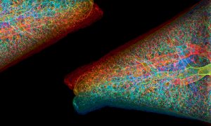
Putting Cryptococcus in context
EMBL’s imaging technology helps researchers gain insights in the fungus’ journey from the lung to the brain.
SCIENCE & TECHNOLOGY2022
sciencescience-technology
Showing results out of

EMBL’s imaging technology helps researchers gain insights in the fungus’ journey from the lung to the brain.
SCIENCE & TECHNOLOGY2022
sciencescience-technology
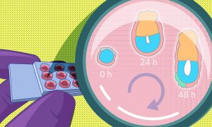
A recent study by EMBL researchers proposes a new method to grow early embryos in the laboratory. With a 3D culture set-up, scientists can closely monitor the changes embryos undergo around the time of implantation.
SCIENCE & TECHNOLOGY2022
sciencescience-technology
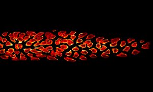
This group of cells represents an interesting example of organ formation where cells simultaneously move and change their shapes in a highly coordinated manner.
SCIENCE & TECHNOLOGY2020
picture-of-the-weekscience-technology
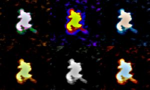
Researchers from EMBL Grenoble have developed a way to visualise large RNAs in 3D using biochemical and structural biology techniques.
SCIENCE & TECHNOLOGY2020
sciencescience-technology
Mature cells can be reprogrammed to pluripotency and thus regain the ability to divide and differentiate into specialized cell types. Although these so-called induced pluripotent stem cells (iPS cells) represent a milestone in stem cell research, many of the biochemical processes that underlie…
SCIENCE & TECHNOLOGY2013
sciencescience-technology
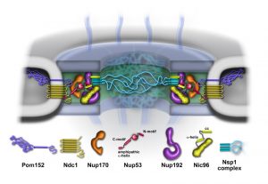
A fungus that lives at extremely high temperatures could help understand structures within our own cells. Scientists at the European Molecular Biology Laboratory (EMBL) and Heidelberg University, both in Heidelberg, Germany, were the first to sequence and analyse the genome of a heat-loving fungus,…
SCIENCE & TECHNOLOGY2011
sciencescience-technology
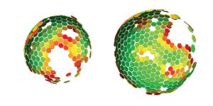
Scientists at the European Molecular Biology Laboratory (EMBL) and the University Clinic Heidelberg, Germany, have produced a three-dimensional reconstruction of HIV (Human Immunodeficiency Virus), which shows the structure of the immature form of the virus at unprecedented detail. Immature HIV is…
SCIENCE & TECHNOLOGY2009
sciencescience-technology
It can be found in all life forms, and serves a multitude of purposes, from energy storage to stress response to bone calcification. This molecular jack-of-all trades is polyphosphate, a long chain of phosphate molecules. Researchers at the European Molecular Biology Laboratory (EMBL) in…
SCIENCE & TECHNOLOGY2009
sciencescience-technology
What at the first sight could be pictures of planets or other cosmic structures are actually microscope images of balls (cysts) of human kidney cells. They were taken by Emmanuel Reynaud, in the group of Ernst Stelzer at the European Molecular Biology Laboratory (EMBL), with a widefield microscope.…
SCIENCE & TECHNOLOGY2008
sciencescience-technology
Researchers at the European Molecular Biology Laboratory (EMBL) have generated a digital zebrafish embryo – the first complete developmental blueprint of a vertebrate. With a newly developed microscope scientists could for the first time track all cells for the first 24 hours in the life of a…
SCIENCE & TECHNOLOGY2008
sciencescience-technology
In the early days of X-ray crystallography obtaining a three-dimensional model of a protein required wire models, screws, bolts and years of tedious calculations by hand. Today macromolecular models are built by computers – thanks to sophisticated software and in particular a package called…
LAB MATTERSSCIENCE & TECHNOLOGY2006
lab-mattersscience-technology

A novel high-tech microscope will be brought to the marketplace, giving laboratories everywhere fascinating new insights into living organisms. EMBLEM Technology Transfer GmbH (EMBLEM), the commercial entity of the European Molecular Biology Laboratory (EMBL), announced today that it has signed a…
CONNECTIONS2005
connections
No results found