29 October 2019
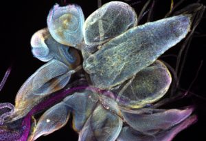
The three bluish blobs shown in the top right corner of this image may not resemble the sphere of noodles that is the human brain, but they are still essential – at least for the fruit fly. This Picture of the Week shows the brain lobes of Drosophila. It’s an insect so tiny and so […]
SCIENCE & TECHNOLOGY
2019
picture-of-the-weekscience-technology
7 May 2019
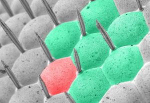
The hexagons visible in this Picture of the Week are the eyes of an ordinary housefly, visualised with a scanning electron microscope. Former staff member Anna Steyer, who captured this brilliant image, has coloured seven of the receptor areas of the eye to create a stylised version…
LAB MATTERS
2019
lab-matterspicture-of-the-week
3 June 2012
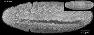
“This video shows a fruit fly embryo from when it was about two-and-a-half hours old until it walked away from the microscope as a larva, 20 hours later,” says Lars Hufnagel, from the European Molecular Biology Laboratory (EMBL) in Heidelberg, Germany. “It shows all the hallmarks of fruit fly…
SCIENCE & TECHNOLOGY
2012
sciencescience-technology
2 February 2012
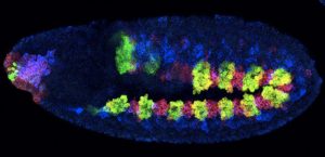
If you wanted to draw your family tree, you could start by searching for people who share your surname. Cells, of course, don’t have surnames, but scientists at the European Molecular Biology Laboratory (EMBL) in Heidelberg, Germany, have found that genetic switches called enhancers, and the…
SCIENCE & TECHNOLOGY
2012
sciencescience-technology
25 June 2009
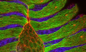
Researchers at the European Molecular Biology Laboratory (EMBL) in Heidelberg, Germany, came a step closer to understanding how cells close gaps not only during embryonic development but also during wound healing. Their study, published this week in the journal Cell, uncovers a fundamental…
SCIENCE & TECHNOLOGY
2009
sciencescience-technology


