No results found
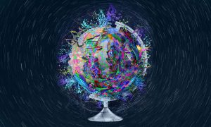
Seeing is understanding
Imaging lets us observe biology in action – it makes visible the hidden processes of life. From its founding, EMBL has been a centre of breakthroughs and developments in bioimaging, and it continues to play a pioneering role in this field today.
SCIENCE & TECHNOLOGY2024
science-technology
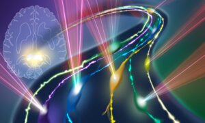
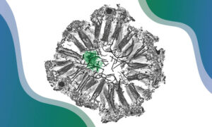
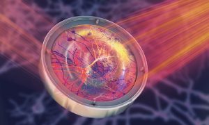
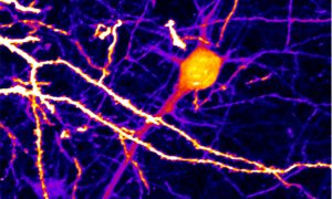
Follow the cellular road
An AI-enhanced advanced microscopy approach offers promise in better understanding glioblastomas, one of the deadliest brain cancers.
SCIENCE & TECHNOLOGY2024
science-technology

Impact of access to imaging technologies on scientific achievements
Baubak Bajoghli, Director of Austrian Bioimaging/CMI, discusses his passion for imaging and his work straddling basic and applied research in biology.
EMBLetc2023
Five takeaways from ‘Seeing is believing’ in 2023
A recent EMBO | EMBL symposium brought together leading developers of imaging methods with cutting-edge applications that illustrate how imaging can answer biological questions.
SCIENCE & TECHNOLOGY2023
eventsscience-technology

Five decades of EMBL visits
Structural biologist Shlomo Trachtenberg has made research trips to EMBL from Israel since the late 1970s and reflects on the boost EMBL’s technology provided his research, the ingredients for an ideal research institution, and his ongoing fascination with microscopes.
CONNECTIONSLAB MATTERS2023
connectionslab-matters
Global guidelines to improve the quality of microscopy images in scientific publications
A working group of researchers from the QUAREP-LiMi initiative has developed global guidelines to improve the quality of microscopy data and images published in scientific publications.
SCIENCE & TECHNOLOGY2023
sciencescience-technology
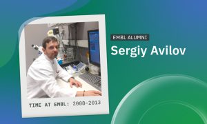
After EMBL: Sergiy Avilov
Ukrainian scientist Sergiy Avilov uses the microscopy skills and scientific network he built at EMBL in his current role heading the Imaging Facility at the Max Planck Institute of Immunobiology and Epigenetics.
PEOPLE & PERSPECTIVES2023
alumnipeople-perspectives
Imaging-based spatial-omics: EMBO Practical Course at EMBL Rome
The first EMBO Practical Course on imaging-based spatial-omics was organised at EMBL Rome to explore the latest techniques to visualise RNA transcripts and proteins in their native tissues.
LAB MATTERSSCIENCE & TECHNOLOGY2023
lab-mattersscience-technology

EMBL and ZEISS enter long-term strategic partnership
Through the collaborative framework agreement, EMBL and ZEISS aim to accelerate the development of imaging technology to advance life science research.
CONNECTIONSLAB MATTERS2023
connectionslab-matters
IMAGINE-ing next-generation technology to help biologists
EMBL researchers and collaborators have begun an impactful innovation-development journey, thanks to a European Commission ‘IMAGINE’ grant.
CONNECTIONSLAB MATTERS2023
connectionslab-matters

Dispensing microscopy expertise
Home to some of Europe’s most cutting-edge tools in molecular biology, EMBL has long shared its expertise and access to these tools through an extensive repertoire of courses, conferences, seminars, and other training. And now included in this mix is a job shadowing programme at EMBL Imaging…
LAB MATTERSSCIENCE & TECHNOLOGY2023
lab-mattersscience-technology

Welcome: Thomas Quail
New group leader Thomas Quail studies the fundamental processes that determine how proteins organise the genome inside a cell.
LAB MATTERSPEOPLE & PERSPECTIVES2023
lab-matterspeople-perspectives
Light-Seq: from images to sequences in context
Researchers have combined advanced light microscopy with next-generation sequencing to create a method to study cells directly in the context of their native tissues
2022
science
A cellular atlas of an entire worm
EMBL scientists and colleagues have developed an interactive atlas of the entire marine worm Platynereis dumerilii in its larval stage. The PlatyBrowser resource combines high-resolution gene expression data with volume electron microscopy images.
SCIENCE & TECHNOLOGY2021
sciencescience-technology
New microscopy technique makes deep in vivo brain imaging possible
Scientists in EMBL’s Prevedel Group have developed a pioneering microscopy technique that allows researchers to observe cells hidden within opaque tissues, such as live neurons embedded deep in the brain.
SCIENCE & TECHNOLOGY2021
sciencescience-technology
Cryo-Electron Microscopy Service Platform supports research on protein transport
Giulia Zanetti from the Institute of Structural and Molecular Biology (ISMB) in London explains how the collaboration with the Cryo-Electron Microscopy Service Platform enabled her group to reveal the structure of protein transport complexes.
LAB MATTERSSCIENCE & TECHNOLOGY2021
lab-mattersscience-technology
Welcome: Liz Duke
The new team leader at EMBL Hamburg talks about her plans to establish biological X-ray imaging and high-throughput tomography.
LAB MATTERSPEOPLE & PERSPECTIVES2021
lab-matterspeople-perspectives
Chan Zuckerberg Initiative recognises EMBL scientists
The Chan Zuckerberg Initiative has recognised four EMBL researchers with their most recent awards, showing how tech trailblazers are integral to advancing science and medicine.
EMBL ANNOUNCEMENTSLAB MATTERS2020
embl-announcementslab-matters

Welcome: Simone Mattei
The new team leader offering services in electron microscopy discusses his hopes and plans for the forthcoming EMBL Imaging Centre
LAB MATTERSPEOPLE & PERSPECTIVES2020
lab-matterspeople-perspectives

Building Euro-BioImaging
EMBL’s Jan Ellenberg reflects on the process of forming a European research infrastructure
LAB MATTERS2019
lab-matters
ATTRACT: funding innovation in imaging
EMBL is a collaborator in four of the projects funded in the first phase of ATTRACT.
EMBL ANNOUNCEMENTSLAB MATTERS2019
embl-announcementslab-matters

A new home for biological images
The BioImage Archive - EMBL-EBI's first dedicated imaging data resource
SCIENCE & TECHNOLOGY2019
sciencescience-technology
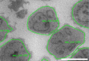
Breaking the bottleneck in imaging data collection
Scientists develop software tools for automated acquisition of electron microscopy data
SCIENCE & TECHNOLOGY2019
sciencescience-technology

Welcome: Santiago Rompani
New EMBL group leader explores what neurobiology can teach us about what it means to be human
PEOPLE & PERSPECTIVES2019
people-perspectivesscience

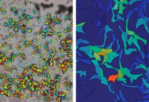
An in silico hope for biology: machine learning
How EMBL scientists are using machine learning to advance biology
LAB MATTERSSCIENCE & TECHNOLOGY2018
lab-mattersscience-technology

Welcome: Jim Swoger
New head of the Mesoscopic Imaging Facility at EMBL Barcelona will help scientists visualise nature
PEOPLE & PERSPECTIVES2018
people-perspectivesscience

Welcome: Simone Köhler
Which of our genes will be passed on to our children? Simone Köhler wants to find out
PEOPLE & PERSPECTIVES2018
people-perspectivesscience

Welcome: Anna Kreshuk
New Heidelberg group leader creates tools to help biologists work faster and better
PEOPLE & PERSPECTIVES2018
people-perspectivesscience
Welcome: Virginie Uhlmann
New EMBL-EBI Group Leader will make sense of bioimaging data
PEOPLE & PERSPECTIVES2018
people-perspectivesscience
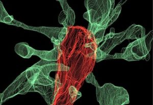
Captured: microglia nibbling on brain synapses
For the first time, EMBL Rome researchers have captured microglia nibbling on brain synapses on film.
SCIENCE & TECHNOLOGY2018
sciencescience-technology

Pan-European imaging infrastructure gains momentum
Europe is uniting to make state-of-the-art imaging technologies accessible to biomedical researchers throughout the continent in a concerted manner. The European Molecular Biology Laboratory (EMBL) and seven countries (Belgium, Finland, Italy, Poland, Slovakia, the Netherlands, the United Kingdom)…
CONNECTIONSLAB MATTERS2014
connectionslab-matters
Become a member of Euro-BioImaging
Euro-BioImaging, the pan-European open access research infrastructure for biological and medical imaging technologies, invites leading European imaging facilities to submit proposals to participate. Applications to become a Euro-BioImaging node will be reviewed by a board of independent…
CONNECTIONSLAB MATTERS2013
connectionslab-matters
Intelligent microscopy
The sight of a researcher sitting at a microscope for hours, painstakingly searching for the right cells, may soon be a thing of the past, thanks to new software created by scientists at the European Molecular Biology Laboratory (EMBL) in Heidelberg, Germany. Presented today in Nature Methods, the…
SCIENCE & TECHNOLOGY2011
sciencescience-technology
Better imaging from bench to bedside
From microscopy to computer tomography (CT) scans, imaging plays an important role in biological and biomedical research, but obtaining high-quality images often requires advanced technology and expertise, and can be costly. Euro-BioImaging, a project which launches its preparatory phase today,…
SCIENCE & TECHNOLOGY2010
sciencescience-technology

The transparent organism
A novel high-tech microscope will be brought to the marketplace, giving laboratories everywhere fascinating new insights into living organisms. EMBLEM Technology Transfer GmbH (EMBLEM), the commercial entity of the European Molecular Biology Laboratory (EMBL), announced today that it has signed a…
CONNECTIONS2005
connections