28 March 2025
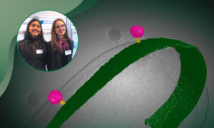
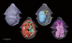
Supported by EMBL services, students Misha Hegde & Mia Maurer have identified a previously unknown bacteriophage that targets Rhizobium rhizogenes, a pathogen harming crops. Using cutting-edge imaging and DNA sequencing at EMBL Heidelberg, they are exploring its potential as a natural alternative…
SCIENCE & TECHNOLOGY
20 March 2025
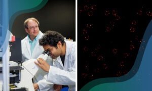
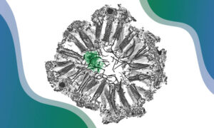
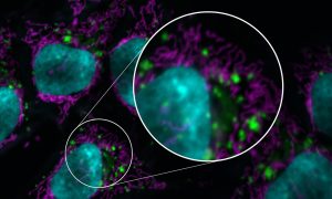
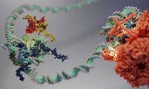
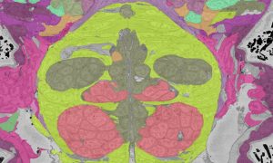
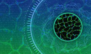
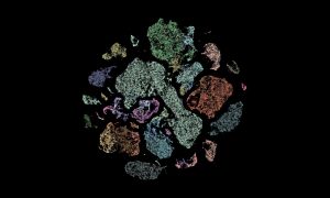
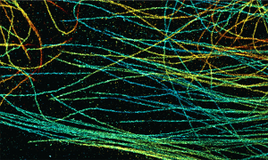
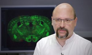
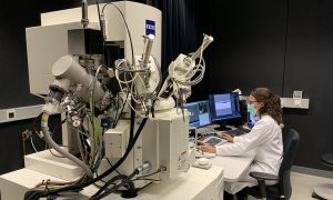
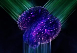
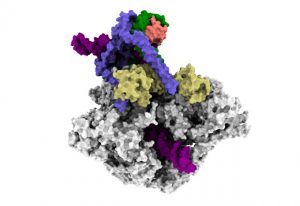
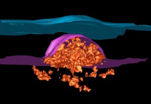
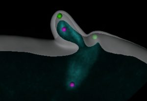
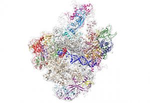
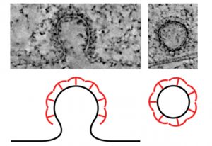
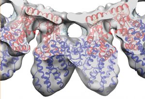
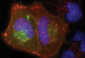
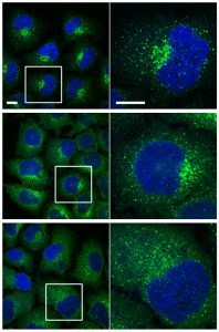
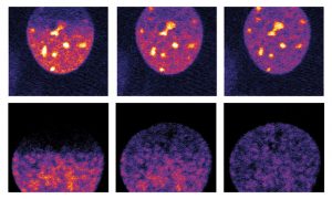
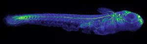
Texas A&M University researchers work with experts from EMBL Imaging Centre to uncover how molecules navigate the nuclear pore complex.
SCIENCE & TECHNOLOGY
20 February 2025
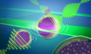
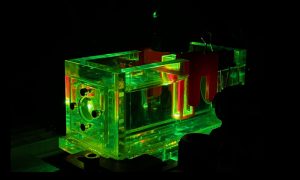
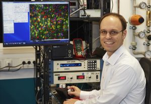
Another EMBL-engineered advance to Brillouin microscopy has significantly widened the aperture to provide quick 3D imaging in real time of light-sensitive samples.
SCIENCE & TECHNOLOGY
17 February 2025
New funding from the Chan Zuckerberg Initiative (CZI) supports two multidisciplinary projects across EMBL’s units and sites to support the development of imaging technologies.
SCIENCE & TECHNOLOGY
17 December 2024
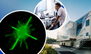
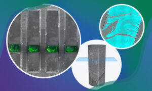
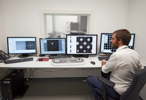
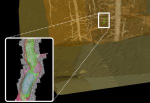
The Prevedel group at EMBL Heidelberg developed a mobile microscope: miniature in scale, fast in sample imaging, and giant in resolution.
SCIENCE & TECHNOLOGY
11 December 2024

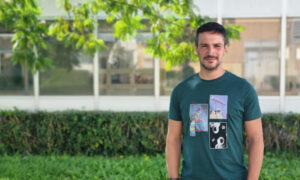
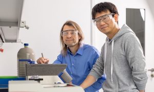
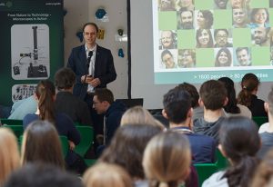
Kristaps Krims-Dāvis spent a month at EMBL Heidelberg getting a crash course in cutting-edge microscopy.
CONNECTIONS
27 November 2024

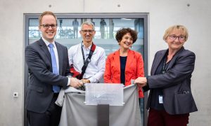
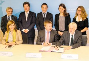
EMBL and Nikon have formally broadened their partnership to accelerate development of next-generation microscopy technologies.
EMBL ANNOUNCEMENTS
17 October 2024
Imaging lets us observe biology in action – it makes visible the hidden processes of life. From its founding, EMBL has been a centre of breakthroughs and developments in bioimaging, and it continues to play a pioneering role in this field today.
SCIENCE & TECHNOLOGY
15 October 2024
Daniele Ancora is an ARISE fellow in the Light Imaging Facility at EMBL Rome. With a background in theoretical physics, he develops algorithms to improve image-based omics technologies. Learn about his interdisciplinary training and his little ‘obsessions’.
PEOPLE & PERSPECTIVES
30 September 2024

EMBL-EBI and colleagues from other 11 institutes commit to pathogen data sharing project.
10 September 2024
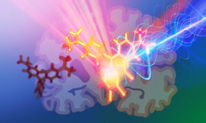
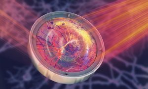
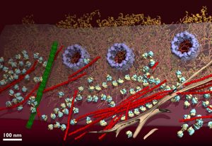
An AI-enhanced advanced microscopy approach offers promise in better understanding glioblastomas, one of the deadliest brain cancers.
SCIENCE & TECHNOLOGY
27 August 2024
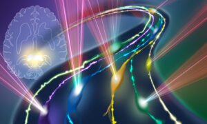
EMBL scientists applied molecular engineering to build photoacoustic probes to label and visualise neurons deep within brain tissue.
SCIENCE & TECHNOLOGY
25 January 2024
Sponges lack muscles and neurons. Yet, they make coordinated movements. Scientists at EMBL Heidelberg have discovered that sponge movement is controlled by an ancient ‘relaxant-inflammatory’ response that is also present in vertebrate blood vessels. The findings shed light on sponge physiology…
SCIENCE & TECHNOLOGY
2024
sciencescience-technology
15 November 2023

Where to start when thinking about the sustainability of informatics projects
15 November 2023

Baubak Bajoghli, Director of Austrian Bioimaging/CMI, discusses his passion for imaging and his work straddling basic and applied research in biology.
EMBLetc
2 November 2023
Users no longer need to download large datasets with the new galleries feature
2023
updates-from-data-resources
27 October 2023
EMBL researchers have created an engineered uterus that allows a closer look at a mouse embryo’s development and its interactions with the uterine environment.
SCIENCE & TECHNOLOGY
2023
picture-of-the-weeksciencescience-technology
5 October 2023
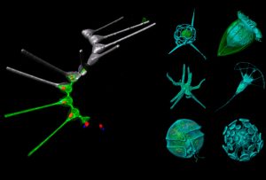
A new method developed by EMBL scientists can help us identify and investigate plankton species in field samples with greater speed, accuracy, and resolution than ever possible before.
SCIENCE & TECHNOLOGY
2023
sciencescience-technology
18 September 2023
A working group of researchers from the QUAREP-LiMi initiative has developed global guidelines to improve the quality of microscopy data and images published in scientific publications.
SCIENCE & TECHNOLOGY
2023
sciencescience-technology
22 August 2023
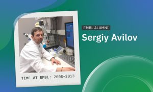
Ukrainian scientist Sergiy Avilov uses the microscopy skills and scientific network he built at EMBL in his current role heading the Imaging Facility at the Max Planck Institute of Immunobiology and Epigenetics.
PEOPLE & PERSPECTIVES
2023
alumnipeople-perspectives
26 July 2023
The first EMBO Practical Course on imaging-based spatial-omics was organised at EMBL Rome to explore the latest techniques to visualise RNA transcripts and proteins in their native tissues.
LAB MATTERSSCIENCE & TECHNOLOGY
2023
lab-mattersscience-technology
21 July 2023
Research from the Eustermann group at EMBL Heidelberg reveals how the packaging of DNA into hexasomes impacts the function of enzymes involved in gene regulation.
SCIENCE & TECHNOLOGY
2023
sciencescience-technology
12 July 2023

Construction begins on EMBL-EBI's new building named after Janet Thornton.
EMBL ANNOUNCEMENTSLAB MATTERS
2023
announcementsembl-announcementslab-matters
10 July 2023
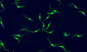
This single-celled organism the size of a dust particle is capable of causing deadly tropical diseases in both humans and livestock –Trypanosoma brucei, in an image by Luciano Dolce from EMBL.
LAB MATTERSSCIENCE & TECHNOLOGY
2023
lab-matterspicture-of-the-weekscience-technology
6 July 2023
Through the collaborative framework agreement, EMBL and ZEISS aim to accelerate the development of imaging technology to advance life science research.
CONNECTIONSLAB MATTERS
2023
connectionslab-matters
31 May 2023
EMBL researchers and collaborators have begun an impactful innovation-development journey, thanks to a European Commission ‘IMAGINE’ grant.
CONNECTIONSLAB MATTERS
2023
connectionslab-matters
16 May 2023

BioImage Archive Team Leader Matthew Hartley shares his experience and tips for people interested in managing a bioinformatics core facility.
2023
technology-and-innovation
15 May 2023
EMBL researchers are pushing the frontiers of big data analysis in biological imaging, allowing scientists to gain a many-layered and multidimensional view of organisms, tissues, and cells in action.
EMBLetc
3 April 2023
Home to some of Europe’s most cutting-edge tools in molecular biology, EMBL has long shared its expertise and access to these tools through an extensive repertoire of courses, conferences, seminars, and other training. And now included in this mix is a job shadowing programme at EMBL Imaging…
LAB MATTERSSCIENCE & TECHNOLOGY
2023
lab-mattersscience-technology
30 March 2023
A new microscope built by EMBL researchers, based on Brillouin scattering principles, allows scientists to observe the dynamics of mechanical properties inside developing embryos in real time.
LAB MATTERSSCIENCE & TECHNOLOGY
2023
lab-matterssciencescience-technology
21 March 2023

New group leader Thomas Quail studies the fundamental processes that determine how proteins organise the genome inside a cell.
LAB MATTERSPEOPLE & PERSPECTIVES
2023
lab-matterspeople-perspectives
3 March 2023
EMBL’s French site was highlighted in a short film presenting its expertise in structural biology research and services.
CONNECTIONSLAB MATTERS
2023
connectionslab-matters
23 February 2023
Researchers in the Prevedel Group use photoacoustic spectroscopy setup to test and optimise probes before their usage in mouse neuroscience.
LAB MATTERSSCIENCE & TECHNOLOGY
2023
lab-matterspicture-of-the-weekscience-technology
22 February 2023

Jo McEntyre talks about data services, open data and a new era for research assessment.
LAB MATTERSPEOPLE & PERSPECTIVES
2023
lab-matterspeople-perspectivesperspectives
11 February 2023
A recent student visitor shares her impressions from visiting EMBL’s Vincent group as we recognise International Day of Women and Girls in Science.
LAB MATTERS
19 January 2023
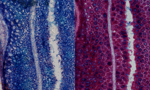
Mucus present in the mouse colon can be visualised using Alcian blue staining, as imaged here by EMBL predoctoral fellow Linda Decker.
LAB MATTERSSCIENCE & TECHNOLOGY
2023
lab-matterspicture-of-the-weekscience-technology
9 November 2022
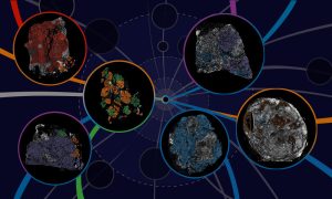
Researchers have created a tool that maps in previously unseen detail how breast cancer develops and spreads.
2022
research-highlightsscience
2 November 2022
Researchers across EMBL are helping to make artificial intelligence (AI) models for bioimaging analysis interoperable and openly available to the scientific community.
2022
announcementsscience
25 October 2022
NIH BRAIN Initiative to fund brain atlases, network coordination, and knowledge sharing to explore brain function research.
2022
announcementsscience
13 October 2022

The Royal Microscopical Society awarded Ardan Patwardhan and Wim Hagen with Scientific Achievement Award
EMBL ANNOUNCEMENTSLAB MATTERS
2022
embl-announcementslab-matters
12 October 2022
Visit of delegation from the Ruđer Bošković Institute to EMBL Heidelberg marks a new chapter in scientific and institutional cooperation
CONNECTIONS
10 October 2022
Researchers have combined advanced light microscopy with next-generation sequencing to create a method to study cells directly in the context of their native tissues
29 September 2022

The Royal Microscopical Society recognises Ardan Patwardhan’s contributions to the field of electron microscopy.
16 September 2022
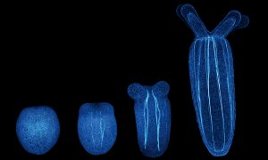
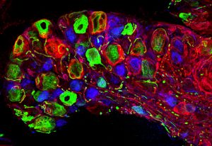
The latest research from EMBL’s Ikmi group employs interdisciplinary approaches to show how sea anemone ‘exercise’ changes their developing size and shape, uncovering an intimate relationship between behaviour and body development
SCIENCE & TECHNOLOGY
2022
sciencescience-technology
24 August 2022
EMBL’s imaging centre makes advanced microscopy technologies accessible to the international scientific community.
LAB MATTERS
2022
lab-matterspicture-of-the-week
5 August 2022
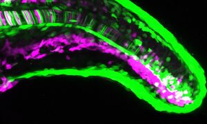
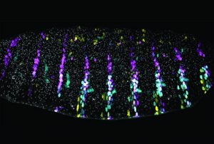
How do gene expression patterns result in the generation of different cell types? Scientists at EMBL Heidelberg used the zebrafish notochord to find out.
SCIENCE & TECHNOLOGY
2022
sciencescience-technology
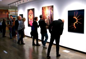
5 July 2022
Creating a cutting-edge facility for the global life science community doesn't happen overnight. We spoke to some of those who worked to turn this dream into a reality.
CONNECTIONS
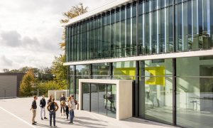
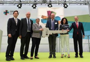
1 July 2022
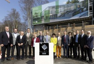
Representatives from politics, industry and academia attended the inauguration ceremony at EMBL Heidelberg
EMBL ANNOUNCEMENTS
2022
embl-announcementsevents
5 May 2022
EMBL’s first Imaging Centre Symposium will occur onsite at EMBL and include tours of the new Imaging Centre on 31 May, introducing participants to the facility and its staff and featuring talks on the rapid developments in imaging technologies that have led to notable biological and medical…
EMBL ANNOUNCEMENTS
2022
embl-announcementsevents
16 March 2022
EMBL’s imaging technology helps researchers gain insights in the fungus’ journey from the lung to the brain.
SCIENCE & TECHNOLOGY
2022
sciencescience-technology
24 January 2022
The new EMBL Imaging Centre held its first on-site training workshop, introducing undergraduate students to the basics of volume electron microscopy. This marks the first of many opportunities to aid capacity-building in imaging techniques in Europe.
LAB MATTERS
3 December 2021
The new EMBL Imaging Centre has announced its first open user call, and new project evaluation process and training opportunities.
EMBL ANNOUNCEMENTSLAB MATTERS
2021
embl-announcementslab-matters
5 October 2021
EMBL scientists and colleagues have developed an interactive atlas of the entire marine worm Platynereis dumerilii in its larval stage. The PlatyBrowser resource combines high-resolution gene expression data with volume electron microscopy images.
SCIENCE & TECHNOLOGY
2021
sciencescience-technology
30 September 2021
Scientists in EMBL’s Prevedel Group have developed a pioneering microscopy technique that allows researchers to observe cells hidden within opaque tissues, such as live neurons embedded deep in the brain.
SCIENCE & TECHNOLOGY
2021
sciencescience-technology
23 September 2021
Some of the most amazing creatures live in the deep blue sea. The Mesoscopic Imaging Facility (MIF) at EMBL Barcelona was recently involved in studying one unique feature of the octopus: the ephemeral structures on the surface of their skin called Kölliker’s organs.
SCIENCE & TECHNOLOGY
2021
sciencescience-technology
10 September 2021
Packaged for simple installation and free use, the novel method DECODE enables researchers to reduce imaging times and increase localisation density in single-molecule localisation microscopy (SMLM).
SCIENCE & TECHNOLOGY
2021
sciencescience-technology
6 July 2021
The EMBL Imaging Centre is preparing for external user access, after an on-time and on-budget build and handover to the science team.
CONNECTIONSLAB MATTERS
2021
connectionslab-matters
23 June 2021

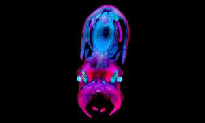
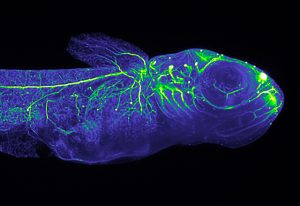
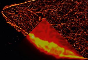
EMBL PhD student Anniek Stokkermans captured this side view of a Nematostella vectensis larva during this transition, using instrumentation in the Advanced Light Microscopy Facility at EMBL Heidelberg.
SCIENCE & TECHNOLOGY
2021
picture-of-the-weekscience-technology
17 June 2021
EMBL group leaders Julia Mahamid, Anna Kreshuk & Jonas Ries awarded Chan Zuckerberg Initiative grant to advance what we see inside cells.
LAB MATTERSPEOPLE & PERSPECTIVES
2021
lab-matterspeople-perspectives
8 June 2021
At EMBL, we have many dream teams – groups of individuals who support each other, innovate, and work together. One of those dream teams bridges two core facilities at EMBL Rome.
SCIENCE & TECHNOLOGY
2021
picture-of-the-weekscience-technology
18 May 2021
The EMBL Picture of the Week features a series of Jurkat T cells during different stages of the activation process.
SCIENCE & TECHNOLOGY
2021
picture-of-the-weekscience-technology
7 April 2021

The challenges and opportunities when setting up a global archive for bioimages
LAB MATTERSPEOPLE & PERSPECTIVES
2021
lab-matterspeople-perspectives
2 December 2020
The Chan Zuckerberg Initiative has recognised four EMBL researchers with their most recent awards, showing how tech trailblazers are integral to advancing science and medicine.
EMBL ANNOUNCEMENTSLAB MATTERS
2020
embl-announcementslab-matters
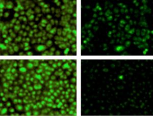
27 October 2020
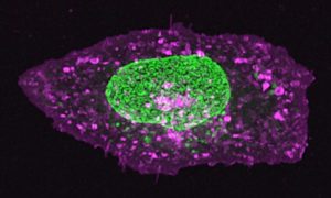
The nucleus of this cell fluoresces in bright green thanks to GFP-labelled nucleoporin proteins. EMBL scientists use engineered nucleoporins as 3D reference standards to improve super-resolution microscopy.
SCIENCE & TECHNOLOGY
2020
picture-of-the-weekscience-technology
8 October 2020
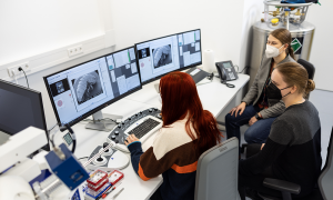
Research facilities play a crucial role in the advancement of science by supporting scientists with specialised expertise and state-of-the-art equipment. The Microscopy Facility at EMBL Rome exemplifies this role by making a wide variety of light microscopy technologies available to its researchers…
LAB MATTERSSCIENCE & TECHNOLOGY
2020
lab-mattersscience-technology
30 April 2020
EMBL electron microscopy specialists collaborate with researchers from Heidelberg University Hospital to understand the changes occurring in cell structures upon SARS-CoV-2 infection.
SCIENCE & TECHNOLOGY
2020
sciencescience-technology
17 April 2020

EMBL Heidelberg reopens the Cryo-Electron Microscopy Service Platform to support coronavirus structural biology research.
CONNECTIONS
7 April 2020
EMBL is all about exciting science, through which we aim to achieve a fundamental understanding of biological processes.
SCIENCE & TECHNOLOGY
2020
picture-of-the-weekscience-technology
17 December 2019
EMBL’s Jan Ellenberg reflects on the process of forming a European research infrastructure
LAB MATTERS
15 November 2019
EMBL is a collaborator in four of the projects funded in the first phase of ATTRACT.
EMBL ANNOUNCEMENTSLAB MATTERS
2019
embl-announcementslab-matters
14 November 2019
EMBL researchers have published two new studies involving the nuclear pore complex
SCIENCE & TECHNOLOGY
2019
sciencescience-technology
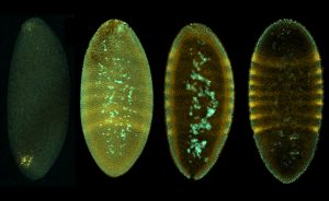
24 July 2019
Enhancers in Drosophila embryos gather together to preserve phenotypes under stressful conditions
SCIENCE & TECHNOLOGY
2019
sciencescience-technology
5 July 2019
EMBL alumna Jennifer Deegan built a prize-winning system for photographing ferns
EMBL ANNOUNCEMENTS
2019
alumniembl-announcements
1 July 2019
A conversation about art-science collaborations and the importance of drawing in biology.
PEOPLE & PERSPECTIVES
2019
people-perspectivesscience
29 April 2019
A newly developed 3D microscope visualises fast biological processes better than ever.
SCIENCE & TECHNOLOGY
2019
sciencescience-technology
1 April 2019
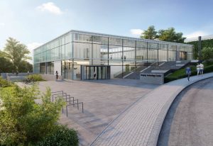
Foundation stone ceremony for world-class high-resolution microscopy centre in Heidelberg
LAB MATTERS
16 November 2018

How EMBL scientists are using machine learning to advance biology
LAB MATTERSSCIENCE & TECHNOLOGY
2018
lab-mattersscience-technology
25 May 2018

EMBL alumna Melina Schuh recognised for excellence in science
PEOPLE & PERSPECTIVES
2018
alumnipeople-perspectives
9 April 2018
Open-source software allows standard microscopes to accurately image 3D structures
SCIENCE & TECHNOLOGY
2018
sciencescience-technology
9 April 2018
EMBL scientists count and locate chromosomal proteins during cell duplication
SCIENCE & TECHNOLOGY
2018
sciencescience-technology
12 March 2018
School students build fluorescence microscopes designed by members of the Prevedel group and ELLS
LAB MATTERS
23 February 2018
Access to state-of-the-art microscopes and outstanding expertise
SCIENCE & TECHNOLOGY
2018
sciencescience-technology
17 January 2018
EMBL researchers uncover how a key enzyme that helps cells make new proteins starts its work
SCIENCE & TECHNOLOGY
2018
sciencescience-technology
14 December 2017
EMBL physicist-turned-biologist alumni win 2017 Kendrew and Phillipson awards
PEOPLE & PERSPECTIVES
2017
alumnipeople-perspectives
30 November 2017
New method for 3D imaging microorganisms lends insight into the creatures that inhabit our oceans
SCIENCE & TECHNOLOGY
2017
sciencescience-technology
24 November 2017
The Mahamid group studies meso-scale molecular assemblies in intact cells and model organisms at molecular resolution
PEOPLE & PERSPECTIVES
2017
people-perspectivesscience
16 November 2017

Jacques Dubochet, Nobel laureate and former EMBL group leader, reflects on a key aha moment
PEOPLE & PERSPECTIVES
2017
alumnipeople-perspectives
14 November 2017
Inside the Centre for Structural Systems Biology
SCIENCE & TECHNOLOGY
2017
sciencescience-technology
10 November 2017
EMBL and the European synchrotron ESRF extend their Joint Structural Biology Group
CONNECTIONSLAB MATTERS
2017
connectionslab-matters
10 November 2017
As a new cryo-EM facility is inaugurated, EMBL’s Michael Hons describes his role in the project
SCIENCE & TECHNOLOGY
2017
sciencescience-technology
31 August 2017
German state and federal governments agree funding for microscopy centre at EMBL Heidelberg
LAB MATTERS
31 May 2017
EMBL’s Hiroki Asari investigates how our internal state can change the way our eyes work
SCIENCE & TECHNOLOGY
2017
sciencescience-technology
8 May 2017
Accelerating researchers’ access to next-generation light-sheet microscopy
CONNECTIONSLAB MATTERS
2017
connectionslab-matters
10 April 2017
Spanish government and EMBL sign agreement for new site
EMBL ANNOUNCEMENTSLAB MATTERS
2017
embl-announcementslab-matters
24 March 2017
ERC grantee Edward Lemke shares his vision for the next ten years
SCIENCE & TECHNOLOGY
2017
sciencescience-technology
13 March 2017
ERC grantee Eileen Furlong shares her vision for the next ten years
SCIENCE & TECHNOLOGY
2017
sciencescience-technology
15 December 2016
EMBL scientists use new techniques to describe the architecture of conical HIV capsids
SCIENCE & TECHNOLOGY
2016
sciencescience-technology
29 November 2016
How a team of scientists and artists at EMBL transformed microscopy data into stunning 3D images
LAB MATTERSSCIENCE & TECHNOLOGY
2016
lab-mattersscience-technology
24 November 2016
Ernst Stelzer earns 2016 Lennart Philipson award for advances in light sheet microscopy
PEOPLE & PERSPECTIVES
2016
alumnipeople-perspectives
17 November 2016
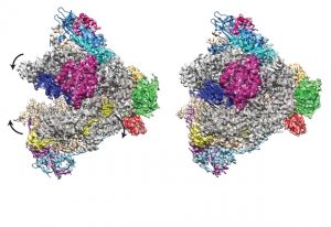
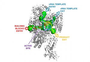
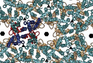
Cryo EM reconstruction of RNA Polymerase I reveals details of how molecule binds and transcribes DNA
SCIENCE & TECHNOLOGY
2016
sciencescience-technology
11 November 2016
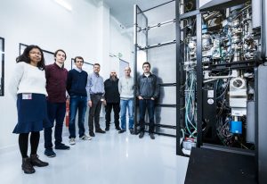
Upgraded server room is ready to take the heat at EMBL Grenoble
LAB MATTERS
27 October 2016
Robert Prevedel develops deep-tissue microscopy for scientists to peer deep inside living organisms
PEOPLE & PERSPECTIVES
2016
people-perspectivesscience
20 October 2016
Participants learn about EMBL’s ocean biodiversity research at the Fall Gala
CONNECTIONS
23 September 2016
Puzzle of nuclear pore formation in growing nuclei solved
SCIENCE & TECHNOLOGY
2016
sciencescience-technology
13 September 2016

A new repository helps identify emerging trends in data-driven science.
LAB MATTERSSCIENCE & TECHNOLOGY
2016
lab-mattersscience-technology
10 August 2016

Storage of pre-made nuclear pores allows for rapid cell division in fruit fly embryos
SCIENCE & TECHNOLOGY
2016
sciencescience-technology
14 July 2016
Study provides insights into workings of new HIV drugs and how virus becomes resistant
SCIENCE & TECHNOLOGY
2016
sciencescience-technology
21 April 2016

How EMBL scientists are discovering and understanding the waves and rhythms inside us
SCIENCE & TECHNOLOGY
2016
sciencescience-technology
21 April 2016

EMBL PhD project puts development in a new light
SCIENCE & TECHNOLOGY
2016
sciencescience-technology
21 March 2016
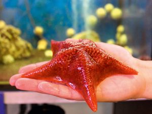
1st real-time video of starfish egg cell eliminating crucial structures, to ensure embryo viability
SCIENCE & TECHNOLOGY
2016
sciencescience-technology
21 March 2016
New technique uses X-rays to find landmarks when combining fluorescence and electron microscopy
SCIENCE & TECHNOLOGY
2016
sciencescience-technology
17 December 2015
From initial development to a start-up company: Selective Plane Illumination Microscopy (SPIM) at EMBL.
SCIENCE & TECHNOLOGY
2015
sciencescience-technology
14 December 2015
New microscope can record the first days of a mouse embryo’s life
SCIENCE & TECHNOLOGY
2015
sciencescience-technology
25 November 2015
CryoEM solves 3D atomic structure of largest and most elusive RNA polymerase.
SCIENCE & TECHNOLOGY
2015
sciencescience-technology
19 November 2015
Using lasers to shed light on how tissues get into shape
SCIENCE & TECHNOLOGY
2015
sciencescience-technology
26 August 2015
"It's like living a review!" Participants of recent super-resolution microscopy course share their highlights
CONNECTIONS
24 August 2015
A journalist who spent six weeks aboard Tara reflects on the expedition’s extraordinary outcomes.
SCIENCE & TECHNOLOGY
2015
sciencescience-technology
18 June 2015
Behaviour of clathrin proteins, crucial for endocytosis, is clarified using new imaging techniques.
SCIENCE & TECHNOLOGY
2015
sciencescience-technology
21 May 2015
Detailed structural study shows distantly related viruses share a common machinery for replication.
SCIENCE & TECHNOLOGY
2015
sciencescience-technology
12 May 2015
Not all embryonic macrophages are the same, and only some are destined to become microglia.
SCIENCE & TECHNOLOGY
2015
sciencescience-technology
7 May 2015
Unveiling the shape of... the 'molecular bin man' – cryoEM helps reveals p62 polymer in 3D.
SCIENCE & TECHNOLOGY
2015
sciencescience-technology
20 April 2015
Ground-breaking microscopy technique gives unprecedented insight into endocytosis.
SCIENCE & TECHNOLOGY
2015
sciencescience-technology
16 March 2015
New fully automated technique enables scientists to chart complex protein networks in living cells.
SCIENCE & TECHNOLOGY
2015
sciencescience-technology
4 March 2015
Combining three different kinds of microscopy to determine how molecules move during endocytosis.
SCIENCE & TECHNOLOGY
2015
sciencescience-technology
25 February 2015
How strong does a spindle need to be? Videos put cell’s chromosome-separating machinery to the test
SCIENCE & TECHNOLOGY
2015
sciencescience-technology
4 February 2015
New microscopy-based method goes beyond gene sequencing, pinpointing the cause of disease.
SCIENCE & TECHNOLOGY
2015
sciencescience-technology
26 January 2015
A brief history of microscopes, from van Leeuwenhoek to Betzig, Hell and Moerner.
SCIENCE & TECHNOLOGY
2015
sciencescience-technology
26 January 2015

From using light to control brain activity to illuminating fruit fly development and mice’s sense of touch
SCIENCE & TECHNOLOGY
2015
sciencescience-technology
23 January 2015

Alumnus Stefan Hell on his 2014 Nobel Prize for Chemistry
LAB MATTERS
16 January 2015

New Christian Boulin Fellowship: 15 awards of up to €1500 for visitors to EMBL’s Core Facilities.
LAB MATTERS
9 January 2015
New group leader Marco Marcia aims to broaden horizons while mapping molecules.
SCIENCE & TECHNOLOGY
2015
sciencescience-technology
18 December 2014
Announcing winners of the John Kendrew Young Scientist Award, and inaugural Lennart Philipson Award.
LAB MATTERS
15 December 2014
Third round of calls for joint research projects between EMBL and Luxembourg in 2015
LAB MATTERS
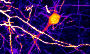
9 December 2014
Unprecedented detail in images of mouse neurons thanks to new SNAP-tagging microscopy technique.
SCIENCE & TECHNOLOGY
2014
sciencescience-technology
3 November 2014
Unprecedented detail on HIV structure continues virus’ string of surprises.
SCIENCE & TECHNOLOGY
2014
sciencescience-technology
20 October 2014
How Nobel-winning work by alumnus Stefan Hell shapes and inspires current EMBL scientists' research.
SCIENCE & TECHNOLOGY
2014
sciencescience-technology
17 October 2014
From anemones to starfish, sea creatures are helping understand development, evolution and more.
SCIENCE & TECHNOLOGY
2014
sciencescience-technology
15 September 2014

Stephen Fuller, from 1981–2000 an EMBL postdoc, group leader then Head of Unit, died on 25 August.
PEOPLE & PERSPECTIVES
2014
alumnipeople-perspectives
12 September 2014
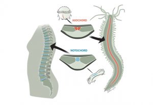
Evolutionary surprise: notochord likely evolved from muscle, earlier than assumed.
SCIENCE & TECHNOLOGY
2014
sciencescience-technology
6 August 2014
How fruit flies beat the cold, plus the value of precisely controlled experiments and detailed analysis
SCIENCE & TECHNOLOGY
2014
sciencescience-technology
1 July 2014

As the Lab turns 40, staff and alumni share 40 things that make EMBL, EMBL
LAB MATTERS
12 September 2013
Scientists at the European Molecular Biology Laboratory (EMBL) in Heidelberg and Regensburg University, both in Germany, and the University of Lisboa, in Portugal, have discovered a promising potential drug target for cystic fibrosis. Their work, published online today in Cell, also uncovers a…
SCIENCE & TECHNOLOGY
2013
sciencescience-technology
11 July 2013
It’s a parent’s nightmare: opening a Lego set and being faced with 500 pieces, but no instructions on how to assemble them into the majestic castle shown on the box. Thanks to a new approach by scientists at the European Molecular Biology Laboratory (EMBL) in Heidelberg, Germany,…
SCIENCE & TECHNOLOGY
2013
sciencescience-technology
3 June 2012
Scientists at the European Molecular Biology Laboratory (EMBL) in Heidelberg, Germany, have conducted the first comprehensive census of human cells’ export workers. In a study published online today in Nature Cell Biology, they found an unexpected variety of genes involved in transporting…
SCIENCE & TECHNOLOGY
2012
sciencescience-technology
7 August 2011
Researchers can now watch molecules move in living cells, literally millisecond by millisecond, thanks to a new microscope developed by scientists at the European Molecular Biology Laboratory (EMBL) in Heidelberg, Germany. Published online today in Nature Biotechnology, the new technique provides…
SCIENCE & TECHNOLOGY
2011
sciencescience-technology
23 January 2011
The sight of a researcher sitting at a microscope for hours, painstakingly searching for the right cells, may soon be a thing of the past, thanks to new software created by scientists at the European Molecular Biology Laboratory (EMBL) in Heidelberg, Germany. Presented today in Nature Methods, the…
SCIENCE & TECHNOLOGY
2011
sciencescience-technology
16 November 2010
The cells in the different parts of this video are always the same (grey), but, like actors using make-up to highlight different facial features, they have fluorescent labels that mark different cellular components in different colours: blue shows the nucleus, yellow shows tubulin (a component of…
SCIENCE & TECHNOLOGY
2010
sciencescience-technology
1 April 2010
Name a human gene, and you’ll find a movie online showing you what happens to cells when it is switched off. This is the resource that researchers at the European Molecular Biology Laboratory (EMBL) in Heidelberg, Germany, and their collaborators in the Mitocheck consortium are making freely…
SCIENCE & TECHNOLOGY
2010
sciencescience-technology
24 February 2009
‘Useless fish with big eyes’. This is what Medaka, the name of the Japanese killifish in the pictures, means in Japan where it originally comes from. While its eyes are undeniably big, the fish has proven remarkably useful for scientists. It is a simple model organism, amenable to…
SCIENCE & TECHNOLOGY
2009
sciencescience-technology
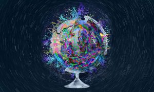
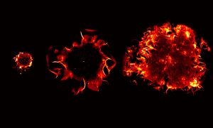
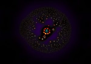
22 October 2008
What at the first sight could be pictures of planets or other cosmic structures are actually microscope images of balls (cysts) of human kidney cells. They were taken by Emmanuel Reynaud, in the group of Ernst Stelzer at the European Molecular Biology Laboratory (EMBL), with a widefield microscope.…
SCIENCE & TECHNOLOGY
2008
sciencescience-technology
4 May 2007
When a cell divides, normally the result is two identical daughter cells. In some cases however, cell division leads to two cells with different properties. This is called asymmetric cell division and plays an important role in embryonic development and the self-renewal of stem cells. Researchers…
SCIENCE & TECHNOLOGY
2007
sciencescience-technology
4 March 2007
The European Molecular Biology Laboratory (EMBL) has developed a new computational tool that makes images obtained with cutting-edge microscopes even sharper. The technological advance and its applications are published in this week’s online issue of the journal Nature Methods. Since the…
SCIENCE & TECHNOLOGY
2007
sciencescience-technology
31 March 2005

A novel high-tech microscope will be brought to the marketplace, giving laboratories everywhere fascinating new insights into living organisms. EMBLEM Technology Transfer GmbH (EMBLEM), the commercial entity of the European Molecular Biology Laboratory (EMBL), announced today that it has signed a…
CONNECTIONS
No results found