Light-Seq: from images to sequences in context
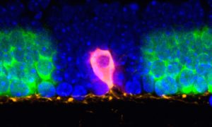
Researchers have combined advanced light microscopy with next-generation sequencing to create a method to study cells directly in the context of their native tissues
2022
science
Showing results out of
Researchers have combined advanced light microscopy with next-generation sequencing to create a method to study cells directly in the context of their native tissues
2022
science
No results found