17 February 2025
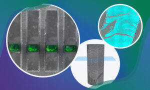
New funding from the Chan Zuckerberg Initiative (CZI) supports two multidisciplinary projects across EMBL’s units and sites to support the development of imaging technologies.
SCIENCE & TECHNOLOGY
15 May 2023
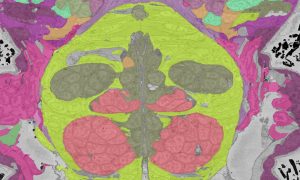
EMBL researchers are pushing the frontiers of big data analysis in biological imaging, allowing scientists to gain a many-layered and multidimensional view of organisms, tissues, and cells in action.
EMBLetc
8 March 2023

EMBL is leading the TREC project: the first pan-European and cross-disciplinary effort to examine life in its natural context.
EMBL ANNOUNCEMENTSLAB MATTERS
2023
embl-announcementslab-matters
31 October 2022
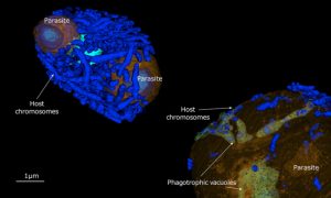
Plankton parasites provide a zombie story perfect for Halloween. While invading single-celled plankton, these parasites devour the cell’s nucleus and hijack metabolism while the organism remains alive.
5 November 2021
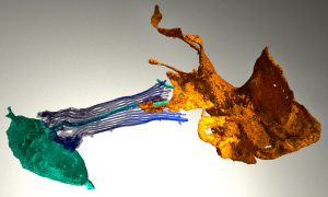
What can sponges tell us about the evolution of the brain? Sponges have the genes involved in neuronal function in higher animals. But if sponges don’t have brains, what is the role of these? EMBL scientists imaged the sponge digestive chamber to find out.
SCIENCE & TECHNOLOGY
2021
sciencescience-technology
5 October 2021

EMBL scientists and colleagues have developed an interactive atlas of the entire marine worm Platynereis dumerilii in its larval stage. The PlatyBrowser resource combines high-resolution gene expression data with volume electron microscopy images.
SCIENCE & TECHNOLOGY
2021
sciencescience-technology
3 June 2021

Under the innovative Planetary Biology research theme, EMBL scientists aim to understand life in the context of its environment.
SCIENCE & TECHNOLOGY
2021
sciencescience-technology
19 January 2021
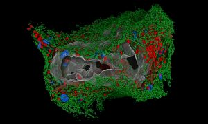
It’s almost a year since the coronavirus outbreak was declared a pandemic, affecting all our lives. While the virus continues its grip on the world, scientists are understanding it better and better, increasing our knowledge about it and opening up new ways to fight it.
SCIENCE & TECHNOLOGY
2021
picture-of-the-weekscience-technology
23 November 2020
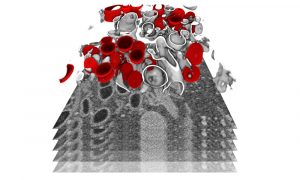
Researchers have studied SARS-CoV-2 replication in cells and obtained detailed insights into the alterations induced in infected cells. This information is essential to guide the development of urgently needed therapeutic strategies for suppressing viral replication and induced pathology.
SCIENCE & TECHNOLOGY
2020
sciencescience-technology
30 April 2020
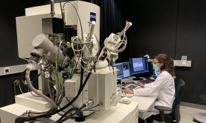
EMBL electron microscopy specialists collaborate with researchers from Heidelberg University Hospital to understand the changes occurring in cell structures upon SARS-CoV-2 infection.
SCIENCE & TECHNOLOGY
2020
sciencescience-technology
28 February 2020
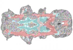
EMBL researchers combine multiple datasets to develop expandable atlas of an entire animal
SCIENCE & TECHNOLOGY
2020
sciencescience-technology
18 February 2020
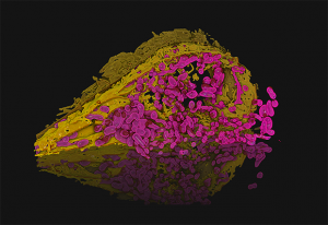
In this image, Julian Hennies from the Schwab Team has reconstructed the 3D structure of a human cell's organelles.
SCIENCE & TECHNOLOGY
2020
picture-of-the-weekscience-technology
21 January 2020
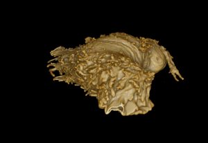
The image shown here is a 3D-rendering created by Julian Hennies from the Schwab team.
SCIENCE & TECHNOLOGY
2020
picture-of-the-weekscience-technology
15 May 2019
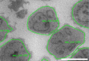
Scientists develop software tools for automated acquisition of electron microscopy data
SCIENCE & TECHNOLOGY
2019
sciencescience-technology
26 March 2018
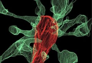
For the first time, EMBL Rome researchers have captured microglia nibbling on brain synapses on film.
SCIENCE & TECHNOLOGY
2018
sciencescience-technology
10 August 2016

Storage of pre-made nuclear pores allows for rapid cell division in fruit fly embryos
SCIENCE & TECHNOLOGY
2016
sciencescience-technology
21 March 2016
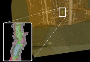
New technique uses X-rays to find landmarks when combining fluorescence and electron microscopy
SCIENCE & TECHNOLOGY
2016
sciencescience-technology


