
Kristina Djinovic Carugo
Head of EMBL Grenoble
ORCID: 0000-0003-0252-2972
EditScientists at EMBL Grenoble focus on integrated structural biology research, and on developing state-of-the art instrumentation, methods and services
Activities at EMBL Grenoble focus on integrated structural biology research, and on developing state-of-the art instrumentation, methods and services. The 3D structure of a biological molecule can tell you a lot about what that molecule does – and how its biological activity might be blocked or altered, for example to treat a disease.
Scientists at EMBL Grenoble determine 3D structures of human and viral proteins to understand how they interact with the nucleic acids DNA and RNA. To do so, they work closely with instrumentation developers and colleagues across the European Photon and Neutron (EPN) science campus to obtain the best possible data from synchrotron X-ray diffraction or cryo-electron microscopy experiments.
EMBL Grenoble shares the European Photon and Neutron (EPN) science campus with the European Synchrotron Radiation Facility (ESRF), the Institut Laue-Langevin (ILL, Europe’s high flux neutron source), and the French Institut de Biologie Structurale (IBS).
The four institutes join forces in the Partnership for Structural Biology (PSB), which provides a uniquely comprehensive range of state-of-the-art structural biology platforms for sample production, sample characterisation and structure determination for both in house research and external users.
Scientists at EMBL Grenoble have, via the PSB, access to a wide range of techniques, including molecular biology and protein expression, biophysical instrumentation, negative stain and cryo-electron microscopy, isotope labelling, nuclear magnetic resonance, neutron scattering, X-ray crystallography, and small angle scattering and imaging.
MBL Grenoble’s researchers focus mainly on RNA biology and infection biology, in particular on the structural molecular biology of protein-RNA complexes involved in cellular gene expression and host-pathogen interactions. The latter includes work on important human viral, parasite and bacterial pathogens. The work typically involves structure determination by X-ray crystallography or cryo-electron microscopy and associated functional studies. Recent highlights include: the structure and function of long non-coding RNAs, the structure of the integrator complex, the determination of the mechanism of action of secreted Legionella effector proteins, and the structural and mechanistic analysis of the transcription/replication machines of influenza virus and lassa virus.
A cornerstone of EMBL Grenoble’s activities is the close interaction with the ESRF, which runs the Extremely Brilliant Source (EBS), the world’s first fourth generation of synchrotron. Through the Joint Structural Biology Group (JSBG), EMBL staff collaborate with the ESRF in building and operating state-of-the-art X-ray beamlines, developing associated instrumentation and techniques, and providing expert help to visitors.
EMBL Grenoble also operates the High Throughput Crystallization Facility (HTX lab), which integrates protein crystallisation into efficient structure determination pipelines. This includes the development of the concept of Online Crystallography, a fully automated and remote controlled pipeline combining automated crystal mounting using the CrystalDirect technology and the CRIMS software. The HTX Facility also promotes structure-guided drug design, through automated facilities for ligand and fragment screening. These platforms are available to external users under the EU funded iNEXT-Discovery project and Instruct-ERIC.
A state-of-the-art Eukaryotic Expression Facility is also available at EMBL Grenoble, which features expression of multiprotein complexes in insect and mammalian cells.
EMBL Grenoble has its own in house Glacios cryo-electron microscope for screening and data collection and participates with the other PSB institutes in running a Krios microscope installed at the ESRF for external users.
Head of EMBL Grenoble
ORCID: 0000-0003-0252-2972
EditWith specialist research groups and teams in both scientific areas research at EMBL Grenoble outstation focuses on structural biology and molecular cell biology. In addition, a number of technology-focused instrumentation teams provide an invaluable resource of technical know how and support to aid the scientific community in the structural biology realm.

Structural biology of ubiquitin signalling
Edit
Structure and assembly mechanisms of sarcomeric cytoskeleton
Edit
Structure and function of RNA-protein complexes
Edit
Structural biology of macromolecular protein-RNA complexes
Edit
Structure and function of lncRNA-protein complexes regulating development and stress responses
Edit
High-throughput crystallisation laboratory
Edit
Synchrotron Crystallography Team
Edit
Multi-scale imaging for evolutionary cell biology
Edit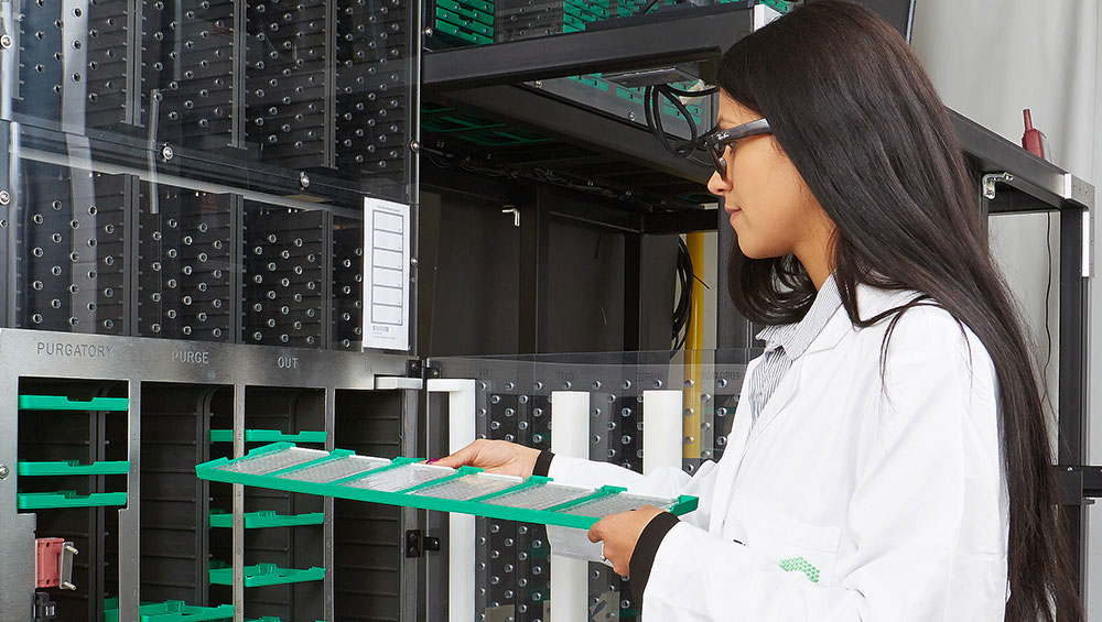
Advancing molecular biology research to study life in context

Research groups at EMBL are organised into nine units spanning six European sites

Explore our latest vacancies and sign up for job alerts to get notified when something suitable comes up